Difference between revisions of "Pseudoepitheliomatous hyperplasia"
Jump to navigation
Jump to search
| (5 intermediate revisions by the same user not shown) | |||
| Line 1: | Line 1: | ||
'''Pseudoepitheliomatous hyperplasia''' is mimic | '''Pseudoepitheliomatous hyperplasia''', abbreviated '''PEH''', is a benign reactive change of squamous mucosa that can mimic [[squamous cell carcinoma]]. | ||
==General== | |||
*Benign. | |||
*Not common. | |||
It is seen in: | It is seen in: | ||
| Line 6: | Line 10: | ||
#[[Granular cell tumour]]. | #[[Granular cell tumour]]. | ||
#Adjacent to an ulcer. | #Adjacent to an ulcer. | ||
#*May form after an injury to the skin.<ref name=pmid17244318>{{Cite journal | last1 = Fu | first1 = X. | last2 = Jiang | first2 = D. | last3 = Chen | first3 = W. | last4 = Sun Bs | first4 = T. | last5 = Sheng | first5 = Z. | title = Pseudoepitheliomatous hyperplasia formation after skin injury. | journal = Wound Repair Regen | volume = 15 | issue = 1 | pages = 39-46 | month = | year = | doi = 10.1111/j.1524-475X.2006.00183.x | PMID = 17244318 }}</ref> | |||
==Microscopic== | |||
Features:<ref name=pmid17614808>{{Cite journal | last1 = Cui | first1 = W. | last2 = McGregor | first2 = DH. | last3 = Stark | first3 = SP. | last4 = Ulusarac | first4 = O. | last5 = Mathur | first5 = SC. | title = Pseudoepitheliomatous hyperplasia - an unusual reaction following tattoo: report of a case and review of the literature. | journal = Int J Dermatol | volume = 46 | issue = 7 | pages = 743-5 | month = Jul | year = 2007 | doi = 10.1111/j.1365-4632.2007.03150.x | PMID = 17614808 }}</ref> | |||
*Acanthosis (epidermal thickening) - irregular .<ref name=pmid21399447>{{Cite journal | last1 = Zayour | first1 = M. | last2 = Lazova | first2 = R. | title = Pseudoepitheliomatous hyperplasia: a review. | journal = Am J Dermatopathol | volume = 33 | issue = 2 | pages = 112-22; quiz 123-6 | month = Apr | year = 2011 | doi = 10.1097/DAD.0b013e3181fcfb47 | PMID = 21399447 }}</ref> | |||
**Involves both follicles and and non-follicular epidermis. | |||
*Hyperkeratosis - thickening of the stratum corneum. | |||
*Parakeratosis - nuclei in the stratum corneum. | |||
DDx: | |||
*[[Squamous cell carcinoma]]. | |||
* | |||
Images | ===Images=== | ||
*[http://www.pathologyoutlines.com/images/prostate/10_14.jpg Pseudoepitheliomatous hyperplasia (pathologyoutlines.com)].<ref>URL: [http://www.pathologyoutlines.com/topic/penscrotumpenssqhyper.html http://www.pathologyoutlines.com/topic/penscrotumpenssqhyper.html]. Accessed on: 10 November 2012.</ref> | *[http://www.pathologyoutlines.com/images/prostate/10_14.jpg Pseudoepitheliomatous hyperplasia (pathologyoutlines.com)].<ref>URL: [http://www.pathologyoutlines.com/topic/penscrotumpenssqhyper.html http://www.pathologyoutlines.com/topic/penscrotumpenssqhyper.html]. Accessed on: 10 November 2012.</ref> | ||
*[http://www.the-dermatologist.com/sites/default/files/issues/January2012/Fungal%20Figure%204.png Pseudoepitheliomatous hyperplasia (the-dermatologist.com)].<ref>URL: [http://www.the-dermatologist.com/content/treating-rare-fungal-infections-coccidioidomycosis http://www.the-dermatologist.com/content/treating-rare-fungal-infections-coccidioidomycosis]. Accessed on: 10 November 2012.</ref> | *[http://www.the-dermatologist.com/sites/default/files/issues/January2012/Fungal%20Figure%204.png Pseudoepitheliomatous hyperplasia (the-dermatologist.com)].<ref>URL: [http://www.the-dermatologist.com/content/treating-rare-fungal-infections-coccidioidomycosis http://www.the-dermatologist.com/content/treating-rare-fungal-infections-coccidioidomycosis]. Accessed on: 10 November 2012.</ref> | ||
| Line 18: | Line 28: | ||
==References== | ==References== | ||
{{Reflist|1}} | {{Reflist|1}} | ||
[[Category:Stuff]] | [[Category:Stuff]] | ||
[[Category:Diagnosis]] | |||
Latest revision as of 15:54, 24 June 2014
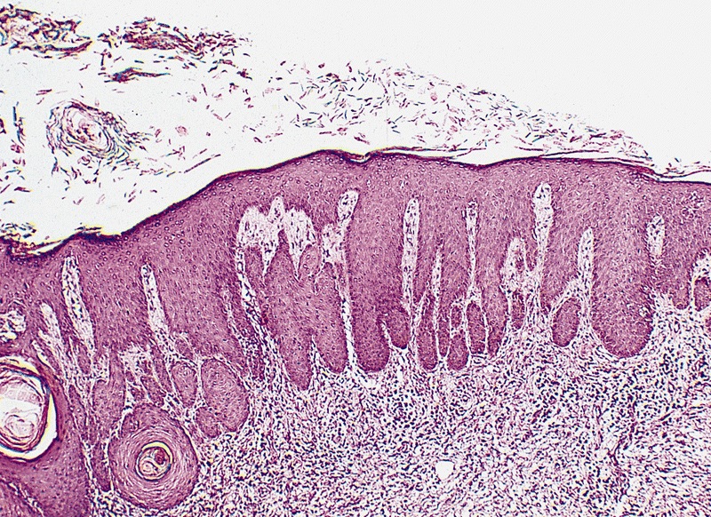
Pseudoepitheliomatous hyperplasia, abbreviated PEH, is a benign reactive change of squamous mucosa that can mimic squamous cell carcinoma.
General
- Benign.
- Not common.
It is seen in:
- Fungal infections.
- Inflammatory papillary hyperplasia.
- Granular cell tumour.
- Adjacent to an ulcer.
- May form after an injury to the skin.[1]
Microscopic
Features:[2]
- Acanthosis (epidermal thickening) - irregular .[3]
- Involves both follicles and and non-follicular epidermis.
- Hyperkeratosis - thickening of the stratum corneum.
- Parakeratosis - nuclei in the stratum corneum.
DDx:
Images
- Pseudoepitheliomatous hyperplasia (pathologyoutlines.com).[4]
- Pseudoepitheliomatous hyperplasia (the-dermatologist.com).[5]
References
- ↑ Fu, X.; Jiang, D.; Chen, W.; Sun Bs, T.; Sheng, Z.. "Pseudoepitheliomatous hyperplasia formation after skin injury.". Wound Repair Regen 15 (1): 39-46. doi:10.1111/j.1524-475X.2006.00183.x. PMID 17244318.
- ↑ Cui, W.; McGregor, DH.; Stark, SP.; Ulusarac, O.; Mathur, SC. (Jul 2007). "Pseudoepitheliomatous hyperplasia - an unusual reaction following tattoo: report of a case and review of the literature.". Int J Dermatol 46 (7): 743-5. doi:10.1111/j.1365-4632.2007.03150.x. PMID 17614808.
- ↑ Zayour, M.; Lazova, R. (Apr 2011). "Pseudoepitheliomatous hyperplasia: a review.". Am J Dermatopathol 33 (2): 112-22; quiz 123-6. doi:10.1097/DAD.0b013e3181fcfb47. PMID 21399447.
- ↑ URL: http://www.pathologyoutlines.com/topic/penscrotumpenssqhyper.html. Accessed on: 10 November 2012.
- ↑ URL: http://www.the-dermatologist.com/content/treating-rare-fungal-infections-coccidioidomycosis. Accessed on: 10 November 2012.