Difference between revisions of "Pleomorphic xanthoastrocytoma"
(split out) |
Jensflorian (talk | contribs) (→Images: APXA) |
||
| (6 intermediate revisions by 2 users not shown) | |||
| Line 1: | Line 1: | ||
'''Pleomorphic xanthoastrocytoma''', abbreviated '''PXA''', is [[neuropathology tumour]] classically associated with seizures. | |||
{{ Infobox diagnosis | |||
| Name = {{PAGENAME}} | |||
| Image = Pleo xantho.jpg | |||
| Width = | |||
| Caption = Pleomorphic xanthoastrocytoma. | |||
| Synonyms = | |||
| Micro = marked nuclear atypia, eosinophilic granular bodies - very common, inflammation (chronic), no necrosis | |||
| Subtypes = | |||
| LMDDx = | |||
| Stains = | |||
| IHC = | |||
| EM = | |||
| Molecular = | |||
| IF = | |||
| Gross = | |||
| Grossing = | |||
| Site = [[brain]] - typical temporal lobe | |||
| Assdx = | |||
| Syndromes = | |||
| Clinicalhx = seizure, children & young adults | |||
| Signs = | |||
| Symptoms = | |||
| Prevalence = | |||
| Bloodwork = | |||
| Rads = | |||
| Endoscopy = | |||
| Prognosis = | |||
| Other = | |||
| ClinDDx = | |||
| Tx = | |||
}} | |||
'''Pleomorphic xanthoastrocytoma''', abbreviated '''PXA''', is [[neuropathology tumour]] classically associated with seizures in children. | |||
==General== | ==General== | ||
Features: | Features: | ||
*Rare (less than 1% of all astrocytic tumors). | |||
*Classically in the temporal lobe in children and young adults. | *Classically in the temporal lobe in children and young adults. | ||
*Associated with seizures. | *Associated with seizures. | ||
*Moderately aggressive (WHO Grade II).<ref name=pmid11465399>{{Cite journal | last1 = Fouladi | first1 = M. | last2 = Jenkins | first2 = J. | last3 = Burger | first3 = P. | last4 = Langston | first4 = J. | last5 = Merchant | first5 = T. | last6 = Heideman | first6 = R. | last7 = Thompson | first7 = S. | last8 = Sanford | first8 = A. | last9 = Kun | first9 = L. | title = Pleomorphic xanthoastrocytoma: favorable outcome after complete surgical resection. | journal = Neuro Oncol | volume = 3 | issue = 3 | pages = 184-92 | month = Jul | year = 2001 | doi = | PMID = 11465399 | URL = http://www.ncbi.nlm.nih.gov/pmc/articles/PMC1920613/pdf/11465399.pdf}}</ref> | *Moderately aggressive (WHO Grade II).<ref name=pmid11465399>{{Cite journal | last1 = Fouladi | first1 = M. | last2 = Jenkins | first2 = J. | last3 = Burger | first3 = P. | last4 = Langston | first4 = J. | last5 = Merchant | first5 = T. | last6 = Heideman | first6 = R. | last7 = Thompson | first7 = S. | last8 = Sanford | first8 = A. | last9 = Kun | first9 = L. | title = Pleomorphic xanthoastrocytoma: favorable outcome after complete surgical resection. | journal = Neuro Oncol | volume = 3 | issue = 3 | pages = 184-92 | month = Jul | year = 2001 | doi = | PMID = 11465399 | URL = http://www.ncbi.nlm.nih.gov/pmc/articles/PMC1920613/pdf/11465399.pdf}}</ref> | ||
*Anaplastic PXA (grade III) - rare. <ref>{{Cite journal | last1 = Louis | first1 = DN. | last2 = Perry | first2 = A. | last3 = Reifenberger | first3 = G. | last4 = von Deimling | first4 = A. | last5 = Figarella-Branger | first5 = D. | last6 = Cavenee | first6 = WK. | last7 = Ohgaki | first7 = H. | last8 = Wiestler | first8 = OD. | last9 = Kleihues | first9 = P. | title = The 2016 World Health Organization Classification of Tumors of the Central Nervous System: a summary. | journal = Acta Neuropathol | volume = 131 | issue = 6 | pages = 803-20 | month = Jun | year = 2016 | doi = 10.1007/s00401-016-1545-1 | PMID = 27157931 }}</ref> | |||
*ICD-O: 9424/3. | |||
==Gross== | ==Gross== | ||
*Temporal lobe - classic. | *Temporal lobe - classic. | ||
*Usually assoc. with the leptomeninges,<ref name=pmid11465399/> i.e. superficial. | *Usually assoc. with the leptomeninges,<ref name=pmid11465399/> i.e. superficial (in up 96%). | ||
<gallery> | |||
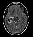
File:405663-PLEOMORPHIC XANTHOASTROCYTOMA.jpg | Typical appearance is one of a superficially situated tumor, here a mural nodule, associated with an underlying cyst (AFIP) | |||
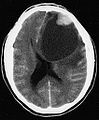
File:Pleomorphic-xanthoastrocytoma-002.jpg | Hemorrhagic pleomorphic xanthoastrocytoma (PXA), MRI (WC/RadsWiki) | |||
</gallery> | |||
==Microscopic== | ==Microscopic== | ||
Features:<ref name=Ref_PBoD8_1333>{{Ref PBoD8|1333}}</ref> | Features:<ref name=Ref_PBoD8_1333>{{Ref PBoD8|1333}}</ref> | ||
* | *Fibrillary background. | ||
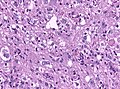
*Large cells with marked nuclear atypia. | |||
*Multinuclear cells possible. | |||
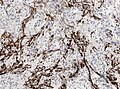
*Reticulin meshwork. | |||
*Lipidized cells. | |||
*Eosinophilic granular bodies - very common.<ref name=pmid11465399/> | *Eosinophilic granular bodies - very common.<ref name=pmid11465399/> | ||
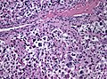
* | *Inflammatory cells (lymophocytic perivascular cuffs). | ||
==Grading== | |||
* Grade II: Mitotic activity is low, no necrosis. | |||
* Grade III anaplastic PXA: more than 5/10 HPF, may have necrosis. | |||
Notes: | Notes: | ||
* | Anaplastic PXA was introduced in the WHO2016 revision as a distinct entity. | ||
* | In the past, the tumor was called ''PXA with anaplastic features''. | ||
DDx: | |||
*[[Subependymal giant cell astrocytoma]]. | |||
*[[Ganglioglioma]]. | |||
*[[Glioblastoma]]. | |||
===Images=== | ===Images=== | ||
<gallery> | |||
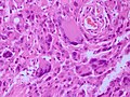
Image:Pleo xantho.jpg | HE smear of a PXA. (WC/AFIP) | |||
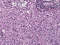
File:PXA HE frozen high mag.jpg | HE intraoperative frozen section of PXA. (WC/jensflorian) | |||
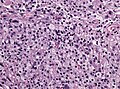
File:PXA HE 40x-2.jpg | The tumor is aptly named due to ist pleomorphic cells. HE, high magnification. (WC/marvin101) | |||
File:PXA HE pleomorphic cells.jpg | The pleomorphic cells must not be confused with a higly anaplastic glioma. HE, intermed magnification.(WC/jensflorian) | |||
File:PXA HE xanthomatous cells.jpg| Focal xanthomatous cells as in the name PXA. HE, intermed magnification.(WC/jensflorian) | |||
File:PXA HE X10.jpg | Perivascular lymphocytic cuffs are not uncommon. HE, intermed-low magnification.(WC/jensflorian) | |||
File:PXA HE x20.jpg | Eosinophilc granular bodies in a PXA. HE, intermed magnification.(WC/jensflorian) | |||
File:PXA Gomori Reticulin Stain.jpg | A delicate meshwork of retiulin fibers in PXA. Gomori, intermed magnification.(WC/jensflorian) | |||
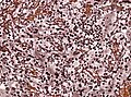
File:PXA GFAP IHC.jpg | GFAP IHC is often heterogenous in PXA. Intermed magnification.(WC/jensflorian) | |||
File:Anaplastic_pxa_histology.jpg| HE of anaplastic PXA. | |||
</gallery> | |||
www: | |||
*[http://path.upmc.edu/cases/case499.html Pleomorphic xanthoastrocytoma - several images (upmc.edu)]. | *[http://path.upmc.edu/cases/case499.html Pleomorphic xanthoastrocytoma - several images (upmc.edu)]. | ||
*[http://path.upmc.edu/cases/case511.html Pleomorphic xanthoastrocytoma with anaplasia - another case - several images (upmc.edu)]. | *[http://path.upmc.edu/cases/case511.html Pleomorphic xanthoastrocytoma with anaplasia - another case - several images (upmc.edu)]. | ||
| Line 35: | Line 102: | ||
==IHC== | ==IHC== | ||
*GFAP +ve. | *GFAP +ve. | ||
*S-100 +ve. | |||
*CD68 +ve. | *CD68 +ve. | ||
*CD34 frequently. | |||
*MAP2+ve and Synapto+ve pleomorphic cells | |||
*MIB-1 usually low. | |||
==Molecular== | |||
* BRAF V600E mutation in 2/3 of the cases<ref>{{Cite journal | last1 = Schindler | first1 = G. | last2 = Capper | first2 = D. | last3 = Meyer | first3 = J. | last4 = Janzarik | first4 = W. | last5 = Omran | first5 = H. | last6 = Herold-Mende | first6 = C. | last7 = Schmieder | first7 = K. | last8 = Wesseling | first8 = P. | last9 = Mawrin | first9 = C. | title = Analysis of BRAF V600E mutation in 1,320 nervous system tumors reveals high mutation frequencies in pleomorphic xanthoastrocytoma, ganglioglioma and extra-cerebellar pilocytic astrocytoma. | journal = Acta Neuropathol | volume = 121 | issue = 3 | pages = 397-405 | month = Mar | year = 2011 | doi = 10.1007/s00401-011-0802-6 | PMID = 21274720 }}</ref> | |||
**(mostly in temporal, reticulin-fiber rich tumors)<ref>{{Cite journal | last1 = Koelsche | first1 = C. | last2 = Sahm | first2 = F. | last3 = Wöhrer | first3 = A. | last4 = Jeibmann | first4 = A. | last5 = Schittenhelm | first5 = J. | last6 = Kohlhof | first6 = P. | last7 = Preusser | first7 = M. | last8 = Romeike | first8 = B. | last9 = Dohmen-Scheufler | first9 = H. | title = BRAF-mutated pleomorphic xanthoastrocytoma is associated with temporal location, reticulin fiber deposition and CD34 expression. | journal = Brain Pathol | volume = 24 | issue = 3 | pages = 221-9 | month = Apr | year = 2014 | doi = 10.1111/bpa.12111 | PMID = 24345274 }}</ref> | |||
==See also== | ==See also== | ||
Latest revision as of 12:38, 11 October 2019
| Pleomorphic xanthoastrocytoma | |
|---|---|
| Diagnosis in short | |
 Pleomorphic xanthoastrocytoma. | |
|
| |
| LM | marked nuclear atypia, eosinophilic granular bodies - very common, inflammation (chronic), no necrosis |
| Site | brain - typical temporal lobe |
|
| |
| Clinical history | seizure, children & young adults |
Pleomorphic xanthoastrocytoma, abbreviated PXA, is neuropathology tumour classically associated with seizures in children.
General
Features:
- Rare (less than 1% of all astrocytic tumors).
- Classically in the temporal lobe in children and young adults.
- Associated with seizures.
- Moderately aggressive (WHO Grade II).[1]
- Anaplastic PXA (grade III) - rare. [2]
- ICD-O: 9424/3.
Gross
- Temporal lobe - classic.
- Usually assoc. with the leptomeninges,[1] i.e. superficial (in up 96%).
Typical appearance is one of a superficially situated tumor, here a mural nodule, associated with an underlying cyst (AFIP)
Hemorrhagic pleomorphic xanthoastrocytoma (PXA), MRI (WC/RadsWiki)


Microscopic
Features:[3]
- Fibrillary background.
- Large cells with marked nuclear atypia.
- Multinuclear cells possible.
- Reticulin meshwork.
- Lipidized cells.
- Eosinophilic granular bodies - very common.[1]
- Inflammatory cells (lymophocytic perivascular cuffs).
Grading
- Grade II: Mitotic activity is low, no necrosis.
- Grade III anaplastic PXA: more than 5/10 HPF, may have necrosis.
Notes: Anaplastic PXA was introduced in the WHO2016 revision as a distinct entity. In the past, the tumor was called PXA with anaplastic features.
DDx:
Images
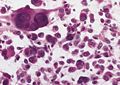
HE smear of a PXA. (WC/AFIP)
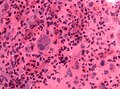
HE intraoperative frozen section of PXA. (WC/jensflorian)
The tumor is aptly named due to ist pleomorphic cells. HE, high magnification. (WC/marvin101)
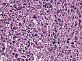
The pleomorphic cells must not be confused with a higly anaplastic glioma. HE, intermed magnification.(WC/jensflorian)
Focal xanthomatous cells as in the name PXA. HE, intermed magnification.(WC/jensflorian)
Perivascular lymphocytic cuffs are not uncommon. HE, intermed-low magnification.(WC/jensflorian)
Eosinophilc granular bodies in a PXA. HE, intermed magnification.(WC/jensflorian)
A delicate meshwork of retiulin fibers in PXA. Gomori, intermed magnification.(WC/jensflorian)
GFAP IHC is often heterogenous in PXA. Intermed magnification.(WC/jensflorian)
HE of anaplastic PXA.


www:
- Pleomorphic xanthoastrocytoma - several images (upmc.edu).
- Pleomorphic xanthoastrocytoma with anaplasia - another case - several images (upmc.edu).
- Pleomorphic xanthoastrocytoma with anaplasia - case 3 - several images (upmc.edu).
- Cerebellar pleomorphic xanthoastrocytoma - case 4 - several image (upmc.edu).
Stains
- Reticulin stain - intercellular, prominent.[4]
Image:
IHC
- GFAP +ve.
- S-100 +ve.
- CD68 +ve.
- CD34 frequently.
- MAP2+ve and Synapto+ve pleomorphic cells
- MIB-1 usually low.
Molecular
See also
References
- ↑ 1.0 1.1 1.2 Fouladi, M.; Jenkins, J.; Burger, P.; Langston, J.; Merchant, T.; Heideman, R.; Thompson, S.; Sanford, A. et al. (Jul 2001). "Pleomorphic xanthoastrocytoma: favorable outcome after complete surgical resection.". Neuro Oncol 3 (3): 184-92. PMID 11465399.
- ↑ Louis, DN.; Perry, A.; Reifenberger, G.; von Deimling, A.; Figarella-Branger, D.; Cavenee, WK.; Ohgaki, H.; Wiestler, OD. et al. (Jun 2016). "The 2016 World Health Organization Classification of Tumors of the Central Nervous System: a summary.". Acta Neuropathol 131 (6): 803-20. doi:10.1007/s00401-016-1545-1. PMID 27157931.
- ↑ Kumar, Vinay; Abbas, Abul K.; Fausto, Nelson; Aster, Jon (2009). Robbins and Cotran pathologic basis of disease (8th ed.). Elsevier Saunders. pp. 1333. ISBN 978-1416031215.
- ↑ 4.0 4.1 Dias-Santagata, D.; Lam, Q.; Vernovsky, K.; Vena, N.; Lennerz, JK.; Borger, DR.; Batchelor, TT.; Ligon, KL. et al. (2011). "BRAF V600E mutations are common in pleomorphic xanthoastrocytoma: diagnostic and therapeutic implications.". PLoS One 6 (3): e17948. doi:10.1371/journal.pone.0017948. PMID 21479234.
- ↑ Schindler, G.; Capper, D.; Meyer, J.; Janzarik, W.; Omran, H.; Herold-Mende, C.; Schmieder, K.; Wesseling, P. et al. (Mar 2011). "Analysis of BRAF V600E mutation in 1,320 nervous system tumors reveals high mutation frequencies in pleomorphic xanthoastrocytoma, ganglioglioma and extra-cerebellar pilocytic astrocytoma.". Acta Neuropathol 121 (3): 397-405. doi:10.1007/s00401-011-0802-6. PMID 21274720.
- ↑ Koelsche, C.; Sahm, F.; Wöhrer, A.; Jeibmann, A.; Schittenhelm, J.; Kohlhof, P.; Preusser, M.; Romeike, B. et al. (Apr 2014). "BRAF-mutated pleomorphic xanthoastrocytoma is associated with temporal location, reticulin fiber deposition and CD34 expression.". Brain Pathol 24 (3): 221-9. doi:10.1111/bpa.12111. PMID 24345274.