Placental meconium
Jump to navigation
Jump to search
The printable version is no longer supported and may have rendering errors. Please update your browser bookmarks and please use the default browser print function instead.
| Placental meconium | |
|---|---|
| Diagnosis in short | |
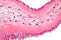
 Meconium-laden macrophages. H&E stain. | |
|
| |
| LM | macrophages with brown fine granular pigment in the amnion +/- chorion |
| LM DDx | hemorrhage |
| Stains | PASD +ve, Prussian blue -ve |
| Gross | green discolourization of fetal membranes |
| Site | placental membranes |
|
| |
| Associated Dx | +/-chorioamnionitis |
| Clinical history | +/-non-reassuring fetal heart rate |
| Prevalence | uncommon |
| Prognosis | benign |
Placental meconium, also known as meconium staining, is associated with fetal distress.
General
- Associated with fetal distress.
- Small amount - at term - is considered to be normal.
Other meconium-related pathology:
Gross
- Fetal membranes with a green discolourization.
Microscopic
Features:[1]
- Meconium histiocytes - key feature.
- Macrophages with brown fine granular pigment.
- Pseudostratified epithelium (amnion) - low power.
- Amnion - columnar morphology (normally cuboidal).
- "Drop-out" of individual amnion cells / loss of individual cells.
Time of meconium passage:[2]
- <1 h - no staining of membranes.
- 1-3 h - amnion is stained.
- >3 h - chorion is stained.
DDx:
- Hemosiderin-laden macrophages.
- This is sorted-out with an iron stain -- see below.
- Chorioamnionitis - apoptotic cells may be mistaken for neutrophils.
Notes:
- The above time course is disputed - in vitro experiments suggest it is considerably longer.[3]
Images
Meconium-laden macrophages - high mag. (WC)
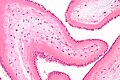
Meconium-laden macrophages - intermed. mag. (WC)

Special stains
- Hemosiderin +ve in hemosiderin-laden macrophages.
- PAS +ve in meconium-laden macrophages.[4]
Useful to differentiate hemosiderin-laden macrophages and meconium laden macrophages:
- Hemosiderin stain -- +ve for old blood.
- Prussian-blue stain = hemosiderin stain.[5]
Notes:
- PAS-D -- +ve in meconium... though may rarely stain hemosiderin.
- Meconium contains bile.[6]
Sign out
PLACENTA, UMBILICAL CORD AND FETAL MEMBRANES, BIRTH:
- FETAL MEMBRANES WITH MECONIUM-LADEN MACROPHAGES, NEGATIVE FOR CHORIOAMNIONITIS.
- THREE VESSEL UMBILICAL CORD WITHIN NORMAL LIMITS.
- PLACENTAL DISC WITH THIRD TRIMESTER VILLI WITHIN NORMAL LIMITS.
COMMENT:
A PAS-D stain and Prussian blue stain were used to confirm the presence of meconium.
Degenerative changes present
PLACENTA, UMBILICAL CORD AND FETAL MEMBRANES, BIRTH:
- FETAL MEMBRANES WITH MECONIUM-LADEN MACROPHAGES AND DEGENERATIVE CHANGES,
  NEGATIVE FOR CHORIOAMNIONITIS.
- THREE VESSEL UMBILICAL CORD WITHIN NORMAL LIMITS.
- PLACENTAL DISC WITH THIRD TRIMESTER VILLI WITHIN NORMAL LIMITS.
Not definite
PLACENTA, UMBILICAL CORD AND FETAL MEMBRANES, BIRTH:
- EARLY CHORIOAMNIONITIS.
- FETAL MEMBRANES WITH FOCAL AMNION CELL DROP-OUT AND RARE PIGMENTED
  CELLS SUGGESTIVE OF MECONIUM.
- THREE VESSEL UMBILICAL CORD WITHIN NORMAL LIMITS.
- PLACENTAL DISC WITH THIRD TRIMESTER VILLI.
See also
References
- ↑ ALS. 6 Febraury 2009.
- ↑ Miller PW, Coen RW, Benirschke K (October 1985). "Dating the time interval from meconium passage to birth". Obstet Gynecol 66 (4): 459–62. PMID 2413412.
- ↑ Funai EF, Labowsky AT, Drewes CE, Kliman HJ (January 2009). "Timing of fetal meconium absorption by amnionic macrophages". Am J Perinatol 26 (1): 93–7. doi:10.1055/s-0028-1103028. PMID 19031358.
- ↑ Povýsil C, Bennett R, Povýsilová V (January 2001). "CD 68 positivity of the so-called meconium corpuscles in human foetal intestine". Cesk Patol 37 (1): 7–9. PMID 11268705.
- ↑ Sienko A, Altshuler G (September 1999). "Meconium-induced umbilical vascular necrosis in abortuses and fetuses: a histopathologic study for cytokines". Obstet Gynecol 94 (3): 415?0. PMID 10472870.
- ↑ Sienko A, Altshuler G (September 1999). "Meconium-induced umbilical vascular necrosis in abortuses and fetuses: a histopathologic study for cytokines". Obstet Gynecol 94 (3): 415?0. PMID 10472870.