Difference between revisions of "Placental meconium"
Jump to navigation
Jump to search
(split-out) |
m (touch) |
||
| (5 intermediate revisions by the same user not shown) | |||
| Line 1: | Line 1: | ||
{{ Infobox diagnosis | {{ Infobox diagnosis | ||
| Name = {{PAGENAME}} | | Name = {{PAGENAME}} | ||
| Image = Meconium-laden_macrophages_high_mag.jpg | | Image = Meconium-laden_macrophages_high_mag.jpg | ||
| Width = | | Width = | ||
| Line 28: | Line 28: | ||
| ClinDDx = | | ClinDDx = | ||
}} | }} | ||
'''Placental meconium''', also known as ''' | '''Placental meconium''', also known as '''meconium staining''', is associated with fetal distress. | ||
==General== | ==General== | ||
| Line 57: | Line 57: | ||
*Hemosiderin-laden macrophages. | *Hemosiderin-laden macrophages. | ||
**This is sorted-out with an iron stain -- see below. | **This is sorted-out with an iron stain -- see below. | ||
*[[Chorioamnionitis]] - apoptotic cells may be mistaken for [[neutrophils]]. | |||
Notes: | Notes: | ||
| Line 66: | Line 67: | ||
Image:Meconium-laden_macrophages_intermed_mag.jpg | Meconium-laden macrophages - intermed. mag. (WC) | Image:Meconium-laden_macrophages_intermed_mag.jpg | Meconium-laden macrophages - intermed. mag. (WC) | ||
</gallery> | </gallery> | ||
==Special stains== | ==Special stains== | ||
*Hemosiderin +ve in hemosiderin-laden macrophages. | *Hemosiderin +ve in hemosiderin-laden macrophages. | ||
| Line 89: | Line 91: | ||
COMMENT: | COMMENT: | ||
A PAS-D stain and Prussian blue stain were used to confirm the presence of meconium. | A PAS-D stain and Prussian blue stain were used to confirm the presence of meconium. | ||
</pre> | |||
===Degenerative changes present=== | |||
<pre> | |||
PLACENTA, UMBILICAL CORD AND FETAL MEMBRANES, BIRTH: | |||
- FETAL MEMBRANES WITH MECONIUM-LADEN MACROPHAGES AND DEGENERATIVE CHANGES, | |||
NEGATIVE FOR CHORIOAMNIONITIS. | |||
- THREE VESSEL UMBILICAL CORD WITHIN NORMAL LIMITS. | |||
- PLACENTAL DISC WITH THIRD TRIMESTER VILLI WITHIN NORMAL LIMITS. | |||
</pre> | </pre> | ||
===Not definite=== | ===Not definite=== | ||
<pre> | <pre> | ||
PLACENTA, UMBILICAL CORD AND FETAL MEMBRANES, | PLACENTA, UMBILICAL CORD AND FETAL MEMBRANES, BIRTH: | ||
- EARLY CHORIOAMNIONITIS. | - EARLY CHORIOAMNIONITIS. | ||
- FETAL MEMBRANES WITH FOCAL AMNION CELL DROP-OUT AND RARE PIGMENTED | - FETAL MEMBRANES WITH FOCAL AMNION CELL DROP-OUT AND RARE PIGMENTED | ||
| Line 104: | Line 115: | ||
*[[Placenta]]. | *[[Placenta]]. | ||
*[[Chorioamnionitis]]. | *[[Chorioamnionitis]]. | ||
*[[Meconium peritonitis]]. | |||
*[[Meconium ileus]]. | |||
==References== | ==References== | ||
Latest revision as of 22:24, 10 January 2015
| Placental meconium | |
|---|---|
| Diagnosis in short | |
 Meconium-laden macrophages. H&E stain. | |
|
| |
| LM | macrophages with brown fine granular pigment in the amnion +/- chorion |
| LM DDx | hemorrhage |
| Stains | PASD +ve, Prussian blue -ve |
| Gross | green discolourization of fetal membranes |
| Site | placental membranes |
|
| |
| Associated Dx | +/-chorioamnionitis |
| Clinical history | +/-non-reassuring fetal heart rate |
| Prevalence | uncommon |
| Prognosis | benign |
Placental meconium, also known as meconium staining, is associated with fetal distress.
General
- Associated with fetal distress.
- Small amount - at term - is considered to be normal.
Other meconium-related pathology:
Gross
- Fetal membranes with a green discolourization.
Microscopic
Features:[1]
- Meconium histiocytes - key feature.
- Macrophages with brown fine granular pigment.
- Pseudostratified epithelium (amnion) - low power.
- Amnion - columnar morphology (normally cuboidal).
- "Drop-out" of individual amnion cells / loss of individual cells.
Time of meconium passage:[2]
- <1 h - no staining of membranes.
- 1-3 h - amnion is stained.
- >3 h - chorion is stained.
DDx:
- Hemosiderin-laden macrophages.
- This is sorted-out with an iron stain -- see below.
- Chorioamnionitis - apoptotic cells may be mistaken for neutrophils.
Notes:
- The above time course is disputed - in vitro experiments suggest it is considerably longer.[3]
Images
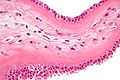
Meconium-laden macrophages - high mag. (WC)
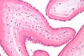
Meconium-laden macrophages - intermed. mag. (WC)

Special stains
- Hemosiderin +ve in hemosiderin-laden macrophages.
- PAS +ve in meconium-laden macrophages.[4]
Useful to differentiate hemosiderin-laden macrophages and meconium laden macrophages:
- Hemosiderin stain -- +ve for old blood.
- Prussian-blue stain = hemosiderin stain.[5]
Notes:
- PAS-D -- +ve in meconium... though may rarely stain hemosiderin.
- Meconium contains bile.[6]
Sign out
PLACENTA, UMBILICAL CORD AND FETAL MEMBRANES, BIRTH:
- FETAL MEMBRANES WITH MECONIUM-LADEN MACROPHAGES, NEGATIVE FOR CHORIOAMNIONITIS.
- THREE VESSEL UMBILICAL CORD WITHIN NORMAL LIMITS.
- PLACENTAL DISC WITH THIRD TRIMESTER VILLI WITHIN NORMAL LIMITS.
COMMENT:
A PAS-D stain and Prussian blue stain were used to confirm the presence of meconium.
Degenerative changes present
PLACENTA, UMBILICAL CORD AND FETAL MEMBRANES, BIRTH:
- FETAL MEMBRANES WITH MECONIUM-LADEN MACROPHAGES AND DEGENERATIVE CHANGES,
  NEGATIVE FOR CHORIOAMNIONITIS.
- THREE VESSEL UMBILICAL CORD WITHIN NORMAL LIMITS.
- PLACENTAL DISC WITH THIRD TRIMESTER VILLI WITHIN NORMAL LIMITS.
Not definite
PLACENTA, UMBILICAL CORD AND FETAL MEMBRANES, BIRTH:
- EARLY CHORIOAMNIONITIS.
- FETAL MEMBRANES WITH FOCAL AMNION CELL DROP-OUT AND RARE PIGMENTED
  CELLS SUGGESTIVE OF MECONIUM.
- THREE VESSEL UMBILICAL CORD WITHIN NORMAL LIMITS.
- PLACENTAL DISC WITH THIRD TRIMESTER VILLI.
See also
References
- ↑ ALS. 6 Febraury 2009.
- ↑ Miller PW, Coen RW, Benirschke K (October 1985). "Dating the time interval from meconium passage to birth". Obstet Gynecol 66 (4): 459–62. PMID 2413412.
- ↑ Funai EF, Labowsky AT, Drewes CE, Kliman HJ (January 2009). "Timing of fetal meconium absorption by amnionic macrophages". Am J Perinatol 26 (1): 93–7. doi:10.1055/s-0028-1103028. PMID 19031358.
- ↑ Povýsil C, Bennett R, Povýsilová V (January 2001). "CD 68 positivity of the so-called meconium corpuscles in human foetal intestine". Cesk Patol 37 (1): 7–9. PMID 11268705.
- ↑ Sienko A, Altshuler G (September 1999). "Meconium-induced umbilical vascular necrosis in abortuses and fetuses: a histopathologic study for cytokines". Obstet Gynecol 94 (3): 415?0. PMID 10472870.
- ↑ Sienko A, Altshuler G (September 1999). "Meconium-induced umbilical vascular necrosis in abortuses and fetuses: a histopathologic study for cytokines". Obstet Gynecol 94 (3): 415?0. PMID 10472870.