Difference between revisions of "Pituitary gland"
Jump to navigation
Jump to search
(create) |
|||
| Line 2: | Line 2: | ||
Divisions:<ref>[http://www.vivo.colostate.edu/hbooks/pathphys/endocrine/hypopit/histo.html http://www.vivo.colostate.edu/hbooks/pathphys/endocrine/hypopit/histo.html]</ref> | Divisions:<ref>[http://www.vivo.colostate.edu/hbooks/pathphys/endocrine/hypopit/histo.html http://www.vivo.colostate.edu/hbooks/pathphys/endocrine/hypopit/histo.html]</ref> | ||
*Anterior pituitary ( | *Anterior pituitary ([[AKA]] adenohypophysis). | ||
*Posterior pituitary ( | *Posterior pituitary (AKA neurohypophysis, neural pituitary). | ||
==Function== | ==Function== | ||
| Line 21: | Line 21: | ||
==Anatomy and histology== | ==Anatomy and histology== | ||
Basic anatomy (simplified):<ref name=bowen>[http://www.vivo.colostate.edu/hbooks/pathphys/endocrine/hypopit/histo_pit.html http://www.vivo.colostate.edu/hbooks/pathphys/endocrine/hypopit/histo_pit.html]</ref> | Basic anatomy (simplified):<ref name=bowen>URL: [http://www.vivo.colostate.edu/hbooks/pathphys/endocrine/hypopit/histo_pit.html http://www.vivo.colostate.edu/hbooks/pathphys/endocrine/hypopit/histo_pit.html]. Accessed on: 31 October 2010.</ref> | ||
*Anterior: | *Anterior: | ||
**Pars distalis. | **Pars distalis. | ||
| Line 31: | Line 31: | ||
*Anterior - Rathke's pouch (roof of mouth). | *Anterior - Rathke's pouch (roof of mouth). | ||
*Posterior - diencephalon (ventral aspect). | *Posterior - diencephalon (ventral aspect). | ||
Image: | |||
*[http://www.ouhsc.edu/histology/Glass%20slides/38_01.jpg Pituitary gland (ouhsc.edu)]. | |||
==Histology== | ==Histology== | ||
Anterior | ===Anterior=== | ||
*Acidophils = red or orange. | *Acidophils (40% of cells) = red or orange. | ||
**GH, PRL. | **GH, PRL. | ||
*Basophils = basophilic (light blue). | *Basophils (10% of cells) = basophilic (light blue). | ||
**TSH, LH, FSH. | **TSH, LH, FSH. | ||
*Chromophobes = | *Chromophobes (50% of cells) = amphophilic (purplish/grey). | ||
Posterior: | Notes: | ||
*The cellular product (i.e. hormone produced) is not strictly correlated with the cell type.<ref name=Ref_PSNP26>{{Ref PSNP|26}}</ref> | |||
===Posterior=== | |||
Features:<ref name=Ref_PSNP26>{{Ref PSNP|26}}</ref> | |||
*Herring bodies - '''key feature'''. | |||
**Eosinophilic axonal dilations filled with lysosomes and neurosecretory granules. | |||
*Less cellular. | *Less cellular. | ||
**Usually more cellular in perivascular location. | |||
Image: [http://www.ouhsc.edu/histology/Glass%20slides/38_09.jpg Herring bodies (ouhsc.edu)]. | |||
==Pituitary adenoma== | |||
Features: | |||
*Loss of fibrous stroma. | |||
==Rathke cleft cyst== | |||
:See ''[[Head and neck pathology]]''. | |||
*Arises from ''intermediate lobe''. | |||
==See also== | ==See also== | ||
Revision as of 01:28, 1 November 2010
The pituitary gland is known as the master gland.
Divisions:[1]
- Anterior pituitary (AKA adenohypophysis).
- Posterior pituitary (AKA neurohypophysis, neural pituitary).
Function
Anterior part:[2]
- Growth hormone (GH).
- Luteinizing hormone (LH)
- Follicle-stimulating hormone (FSH)
- Thyroid stimulating hormone (TSH)
- Adrenocorticotropic hormone (ACTH)
- Prolactin (PRL)
Mnemonic: "Go Look For The Adenoma Please" = GH, LH, FSH, TSH, ACTH, PRL.
Posterior part:[2]
- Oxytocin.
- Antidiuretic hormone (ADH).
Anatomy and histology
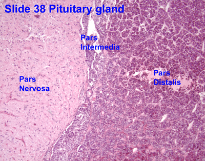
Basic anatomy (simplified):[3]
- Anterior:
- Pars distalis.
- Pars intermedia.
- Posterior:
- Pars nervosa.
Embryological origin:[3]
- Anterior - Rathke's pouch (roof of mouth).
- Posterior - diencephalon (ventral aspect).
Image:
Histology
Anterior
- Acidophils (40% of cells) = red or orange.
- GH, PRL.
- Basophils (10% of cells) = basophilic (light blue).
- TSH, LH, FSH.
- Chromophobes (50% of cells) = amphophilic (purplish/grey).
Notes:
- The cellular product (i.e. hormone produced) is not strictly correlated with the cell type.[4]
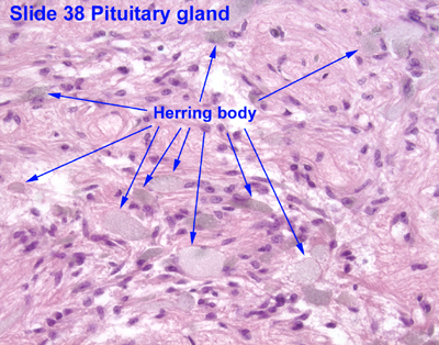
Posterior
Features:[4]
- Herring bodies - key feature.
- Eosinophilic axonal dilations filled with lysosomes and neurosecretory granules.
- Less cellular.
- Usually more cellular in perivascular location.
Image: Herring bodies (ouhsc.edu).
Pituitary adenoma
Features:
- Loss of fibrous stroma.
Rathke cleft cyst
- Arises from intermediate lobe.
See also
References
- ↑ http://www.vivo.colostate.edu/hbooks/pathphys/endocrine/hypopit/histo.html
- ↑ 2.0 2.1 http://users.rcn.com/jkimball.ma.ultranet/BiologyPages/P/Pituitary.html
- ↑ 3.0 3.1 URL: http://www.vivo.colostate.edu/hbooks/pathphys/endocrine/hypopit/histo_pit.html. Accessed on: 31 October 2010.
- ↑ 4.0 4.1 Perry, Arie; Brat, Daniel J. (2010). Practical Surgical Neuropathology: A Diagnostic Approach: A Volume in the Pattern Recognition series (1st ed.). Churchill Livingstone. pp. 26. ISBN 978-0443069826.
External links
- Neuropathology - neuropathologyweb.org.