Difference between revisions of "Pilomatricoma"
Jump to navigation
Jump to search
(split out) |
(more) |
||
| Line 6: | Line 6: | ||
| Micro = "ghost" cells, foreign body-type giant cells | | Micro = "ghost" cells, foreign body-type giant cells | ||
| Subtypes = | | Subtypes = | ||
| LMDDx = | | LMDDx = [[squamous cell carcinoma]], pilomatrix carcinoma, [[basal cell carcinoma]], [[epidermal inclusion cyst]] | ||
| Stains = | | Stains = | ||
| IHC = | | IHC = | ||
| Line 30: | Line 30: | ||
It is also known as '''calcifying epithelioma of Malherbe'''.<ref>{{Ref Derm|387}}</ref> | It is also known as '''calcifying epithelioma of Malherbe'''.<ref>{{Ref Derm|387}}</ref> | ||
==General== | |||
*Benign skin tumour. | *Benign skin tumour. | ||
*Most common solid skin tumour of children.<ref name=emed1058965>URL: [http://emedicine.medscape.com/article/1058965-overview http://emedicine.medscape.com/article/1058965-overview]. Accessed on: 10 September 2011.</ref> | *Most common solid skin tumour of children.<ref name=emed1058965>URL: [http://emedicine.medscape.com/article/1058965-overview http://emedicine.medscape.com/article/1058965-overview]. Accessed on: 10 September 2011.</ref> | ||
| Line 42: | Line 42: | ||
*Surgical excision.<ref name=emed1058965>[http://emedicine.medscape.com/article/1058965-overview http://emedicine.medscape.com/article/1058965-overview]</ref> | *Surgical excision.<ref name=emed1058965>[http://emedicine.medscape.com/article/1058965-overview http://emedicine.medscape.com/article/1058965-overview]</ref> | ||
==Microscopic== | |||
Features:<ref name=emed1058965dx>URL: [http://emedicine.medscape.com/article/1058965-diagnosis http://emedicine.medscape.com/article/1058965-diagnosis]. Accessed on: 10 September 2011.</ref> | Features:<ref name=emed1058965dx>URL: [http://emedicine.medscape.com/article/1058965-diagnosis http://emedicine.medscape.com/article/1058965-diagnosis]. Accessed on: 10 September 2011.</ref> | ||
*Nodular circumscribed lower dermis/subcutaneous adipose lesion; thus, usu. surrounded by connective tissue. | *Nodular circumscribed lower dermis/subcutaneous adipose lesion; thus, usu. surrounded by connective tissue. | ||
| Line 55: | Line 55: | ||
*Keratin a prominent feature on cytology - lots of orange stuff. | *Keratin a prominent feature on cytology - lots of orange stuff. | ||
*May ossify. | *May ossify. | ||
DDx: | DDx: | ||
| Line 67: | Line 61: | ||
*[[Squamous cell carcinoma]]. | *[[Squamous cell carcinoma]]. | ||
*[[Basal cell carcinoma]]. | *[[Basal cell carcinoma]]. | ||
===Images=== | |||
<gallery> | |||
Image:Pilomatrixoma_-_high_mag.jpg | Pilomatrixoma - high mag. (WC/Nephron) | |||
Image:Pilomatrixoma_-_intermed_mag.jpg | Pilomatrixoma - intermed. mag. (WC/Nephron) | |||
</gallery> | |||
www: | |||
*[http://www.bccancer.bc.ca/HPI/CE/cytotechnology/cytosleuthquiz/nongyne/ngcase02.htm Pilomatrixoma - cytology (bccancer.bc.ca)]. | |||
*[http://www.dermrounds.com/photo/1980062:Photo:431 Pilomatrixoma - histology (dermrounds.com)]. | |||
===Sign out=== | ===Sign out=== | ||
Revision as of 15:50, 3 July 2013
| Pilomatricoma | |
|---|---|
| Diagnosis in short | |
 Pilomatricoma. H&E stain. | |
|
| |
| LM | "ghost" cells, foreign body-type giant cells |
| LM DDx | squamous cell carcinoma, pilomatrix carcinoma, basal cell carcinoma, epidermal inclusion cyst |
| Site | skin |
|
| |
| Signs | hard nodule |
| Symptoms | +/-painful |
| Prognosis | benign |
Pilomatricoma is a benign skin lesion that is common in childhood. It may be spelled pilomatrixoma.
It is also known as calcifying epithelioma of Malherbe.[1]
General
- Benign skin tumour.
- Most common solid skin tumour of children.[2]
- CTNNB1 gene mutation important in pathogenesis.[3]
Clinical:
- Hard nodule - calcification.
- +/-Painful.
Treatment:
- Surgical excision.[2]
Microscopic
Features:[4]
- Nodular circumscribed lower dermis/subcutaneous adipose lesion; thus, usu. surrounded by connective tissue.
- Sharpy demarcated island of cells.
- Calcification in 75%.
- Cells:[5]
- Basaloid epithelial cells - have prominent nucleoli.
- Anucleate squamous cells ("ghost cells").
- Giant cell foreign body type granulomas (form in reaction to keratin).
Notes:
- Keratin a prominent feature on cytology - lots of orange stuff.
- May ossify.
DDx:
- Epidermal inclusion cyst.
- Pilomatrix carcinoma - invasive border, cytologic atypia, necrosis.[6]
- Squamous cell carcinoma.
- Basal cell carcinoma.
Images
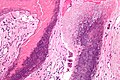
Pilomatrixoma - high mag. (WC/Nephron)
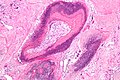
Pilomatrixoma - intermed. mag. (WC/Nephron)

www:
Sign out
SKIN LESION, RIGHT ARM, EXCISION:
- PILOMATRICOMA.
See also
References
- ↑ Busam, Klaus J. (2009). Dermatopathology: A Volume in the Foundations in Diagnostic Pathology Series (1st ed.). Saunders. pp. 387. ISBN 978-0443066542.
- ↑ 2.0 2.1 URL: http://emedicine.medscape.com/article/1058965-overview. Accessed on: 10 September 2011. Cite error: Invalid
<ref>tag; name "emed1058965" defined multiple times with different content - ↑ Mitchell, Richard; Kumar, Vinay; Fausto, Nelson; Abbas, Abul K.; Aster, Jon (2011). Pocket Companion to Robbins & Cotran Pathologic Basis of Disease (8th ed.). Elsevier Saunders. pp. 597. ISBN 978-1416054542.
- ↑ URL: http://emedicine.medscape.com/article/1058965-diagnosis. Accessed on: 10 September 2011.
- ↑ http://www.bccancer.bc.ca/HPI/CE/cytotechnology/cytosleuthquiz/nongyne/ngcase02d.htm
- ↑ Busam, Klaus J. (2009). Dermatopathology: A Volume in the Foundations in Diagnostic Pathology Series (1st ed.). Saunders. pp. 389. ISBN 978-0443066542.