Difference between revisions of "Pilar cyst"
Jump to navigation
Jump to search
(split-out) |
|||
| (8 intermediate revisions by the same user not shown) | |||
| Line 1: | Line 1: | ||
{{ Infobox diagnosis | |||
| Name = {{PAGENAME}} | |||
| Image = Trichilemmal_cyst_-_very_high_mag.jpg | |||
| Width = | |||
| Caption = Pilar cyst. [[H&E stain]]. | |||
| Micro = cyst lining by a stratified squamous epithelium without a granular layer - contains keratin; no significant nuclear atypia; +/-[[granuloma|granulomatous inflammation]] (due to rupture) | |||
| Subtypes = | |||
| LMDDx = [[epidermal inclusion cyst]] | |||
| Stains = | |||
| IHC = | |||
| EM = | |||
| Molecular = | |||
| IF = | |||
| Gross = nodule +/-yellow colour | |||
| Grossing = | |||
| Site = [[skin]] - usu. scalp | |||
| Assdx = | |||
| Syndromes = | |||
| Clinicalhx = | |||
| Signs = | |||
| Symptoms = | |||
| Prevalence = very common | |||
| Bloodwork = | |||
| Rads = | |||
| Endoscopy = | |||
| Prognosis = benign | |||
| Other = | |||
| ClinDDx = other [[skin cysts]] | |||
}} | |||
'''Pilar cyst''', also known as a '''trichilemmal cyst''', is a common benign [[dermal cysts|skin cyst]]. | '''Pilar cyst''', also known as a '''trichilemmal cyst''', is a common benign [[dermal cysts|skin cyst]]. | ||
==General== | ==General== | ||
*Very common. | *Very common. | ||
*Benign. | *Benign.‡ | ||
*The clinical history for these typically says ''[[sebaceous cyst]]''. | |||
Note: | |||
*‡A super rare malignant counter part is described; approximately 40 cases are reported in the english literature.<ref name=pmid22470211>{{Cite journal | last1 = Goyal | first1 = S. | last2 = Jain | first2 = BB. | last3 = Jana | first3 = S. | last4 = Bhattacharya | first4 = SK. | title = Malignant proliferating trichilemmal tumor. | journal = Indian J Dermatol | volume = 57 | issue = 1 | pages = 50-2 | month = Jan | year = 2012 | doi = 10.4103/0019-5154.92679 | PMID = 22470211 }}</ref> | |||
==Gross== | ==Gross== | ||
| Line 18: | Line 51: | ||
DDx: | DDx: | ||
*[[Epidermal cyst]] - has a granular layer. | *[[Epidermal cyst]] - has a granular layer. | ||
*[[Squamous cell carcinoma of the skin|Squamous cell carcinoma]] arising from a pilar cyst. | |||
===Images=== | ===Images=== | ||
<gallery> | <gallery> | ||
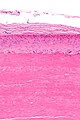
Image:Trichilemmal_cyst_-_intermed_mag.jpg | Trichilemmal cyst - intermed. mag. (WC) | Image:Trichilemmal_cyst_-_intermed_mag.jpg | Trichilemmal cyst - intermed. mag. (WC) | ||
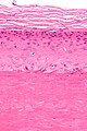
Image:Trichilemmal_cyst_-_high_mag.jpg | Trichilemmal cyst - high mag. (WC) | |||
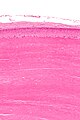
Image:Trichilemmal_cyst_-_very_high_mag.jpg | Trichilemmal cyst - very high mag. (WC) | Image:Trichilemmal_cyst_-_very_high_mag.jpg | Trichilemmal cyst - very high mag. (WC) | ||
</gallery> | </gallery> | ||
| Line 27: | Line 62: | ||
*[http://dermatlas.med.jhmi.edu/derm/indexDisplay.cfm?ImageID=69966366 Trichilemmal cyst (dermatlas.med.jhmi.edu)]. | *[http://dermatlas.med.jhmi.edu/derm/indexDisplay.cfm?ImageID=69966366 Trichilemmal cyst (dermatlas.med.jhmi.edu)]. | ||
*[http://www.flickr.com/photos/40764007@N08/3881846034/in/photostream/ Trichilemmal cyst (flickr.com)]. | *[http://www.flickr.com/photos/40764007@N08/3881846034/in/photostream/ Trichilemmal cyst (flickr.com)]. | ||
==Sign out== | ==Sign out== | ||
<pre> | |||
Scalp (Cyst), Excision: | |||
- Benign trichilemmal cyst (pilar cyst). | |||
</pre> | |||
===Block letters=== | |||
<pre> | <pre> | ||
SCALP (CYST), EXCISION: | SCALP (CYST), EXCISION: | ||
| Line 49: | Line 91: | ||
==References== | ==References== | ||
{{Reflist| | {{Reflist|1}} | ||
[[Category:Diagnosis]] | [[Category:Diagnosis]] | ||
[[Category:Dermal cysts]] | |||
Latest revision as of 13:32, 6 October 2015
| Pilar cyst | |
|---|---|
| Diagnosis in short | |
 Pilar cyst. H&E stain. | |
|
| |
| LM | cyst lining by a stratified squamous epithelium without a granular layer - contains keratin; no significant nuclear atypia; +/-granulomatous inflammation (due to rupture) |
| LM DDx | epidermal inclusion cyst |
| Gross | nodule +/-yellow colour |
| Site | skin - usu. scalp |
|
| |
| Prevalence | very common |
| Prognosis | benign |
| Clin. DDx | other skin cysts |
Pilar cyst, also known as a trichilemmal cyst, is a common benign skin cyst.
General
- Very common.
- Benign.‡
- The clinical history for these typically says sebaceous cyst.
Note:
- ‡A super rare malignant counter part is described; approximately 40 cases are reported in the english literature.[1]
Gross
- Classic location: head ~90%.[2]
Microscopic
Features:[3]
- Keratin.
- Cyst lining:
- Has no granular layer - key feature.
- Keratohyaline granules (as seen in the granular layer) may be seen focally.
- Inner most cyst lining cells are large cells with abundant eosinophilic cytoplasm.
- Has no granular layer - key feature.
DDx:
- Epidermal cyst - has a granular layer.
- Squamous cell carcinoma arising from a pilar cyst.
Images
Trichilemmal cyst - intermed. mag. (WC)
Trichilemmal cyst - high mag. (WC)
Trichilemmal cyst - very high mag. (WC)


www:
Sign out
Scalp (Cyst), Excision:
- Benign trichilemmal cyst (pilar cyst).
Block letters
SCALP (CYST), EXCISION:
- TRICHILEMMAL CYST (PILAR CYST).
SKIN CYST, LEFT FLANK, EXCISION:
- TRICHILEMMAL CYST (PILAR CYST).
SCALP (CYST), ANTERIOR, EXCISION:
- TRICHILEMMAL CYST (PILAR CYST), RUPTURED.
Micro
The sections show a cyst that is lined by squamous epithelium without a granular layer. Focally, keratohyaline granules are seen in the cyst lining cells. The innermost cyst lining cells are large and have abundant eosinophilic cytoplasm. The cyst contains keratin.
See also
References
- ↑ Goyal, S.; Jain, BB.; Jana, S.; Bhattacharya, SK. (Jan 2012). "Malignant proliferating trichilemmal tumor.". Indian J Dermatol 57 (1): 50-2. doi:10.4103/0019-5154.92679. PMID 22470211.
- ↑ URL: http://emedicine.medscape.com/article/1058907-overview. Accessed on: 15 April 2012.
- ↑ Busam, Klaus J. (2009). Dermatopathology: A Volume in the Foundations in Diagnostic Pathology Series (1st ed.). Saunders. pp. 309. ISBN 978-0443066542.