Pediatric gastrointestinal pathology
This article deals with pediatric gastrointestinal pathology. An introduction to pediatric pathology is in the pediatric pathology article.
An overview of (adult) gastrointestinal pathology is in the gastrointestinal pathology article.
Luminal pathology
Microvillous inclusion disease
General
Microscopic
Features:
- Flat mucosa; no villi.
IHC:
- Carcinoembryonic antigen (CEA) +ve.[2]
EM:
- Diagnosis is dependent on electron microscopy.[3]
- Images: MID (gfmer.ch).
Notes:
- Appearance similar to celiac sprue; however, usually lacks the intraepithelial lymphocytic infiltration characteristic of celiac sprue.
Cystic fibrosis
- Abbreviated CF.
General
- Genetic.
- May lead to meconium ileus.
Microscopic (large bowel)
Features:[4]
- Crypt enlargement.
Notes:
- Not intracellular and extracellular accumulation of mucus. (?)
Aganglionosis
- AKA Hirschsprung disease.
General
- Congenital.
- Fixed by surgery.
Pathology:
- Parasympathetic ganglion cells in intramural and submucosal plexuses - not present.[5]
Notes:
- Most common reason for litigation in paediatric pathology.[6]
Microscopic
Features:[5]
- Ganglion cells missing in submucosal plexus and myenteric plexus.
- +/-Submucosal fibrosis.
Stains
- Acetylcholinesterase: abundant, disorganized, nerve fibers.
- CD117. (???)
Images:
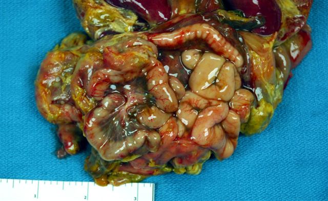
Meconium peritonitis
General
- May be due to a number of causes:
- Aganglionosis (Hirschsprung disease).
- Meconium ileus.
Microscopic
Features:
- Brown granular material - key feature.
- +/-Multinucleated giant cells.
- Inflammatory infiltrate (PMNs, lymphocytes, plasma cells).
Image:
Necrotizing enterocolitis
General
- Disease of the newborn.
- Diagnosed by imaging.
Microscopic
Features:
- Large spaces.
Images:
Pancreatic islet cell hyperplasia
General
- Assoc. with maternal diabetes.
Microscopic
Features:
- Marked size variation of pancreatic islets.
- Normal islets ~ 150 micrometers (diameter). Hyperplastic islets - up to ~500 micrometers (diameter).
Image:
Liver
Neonatal (giant cell) hepatitis
General
- Good prognosis. (???)
Microscopic
Features:
- Hepatocyte enlargement - "ballooning".
- Giant cell formation.
See also
References
- ↑ Müller T, Hess MW, Schiefermeier N, et al. (October 2008). "MYO5B mutations cause microvillus inclusion disease and disrupt epithelial cell polarity". Nat. Genet. 40 (10): 1163–5. doi:10.1038/ng.225. PMID 18724368.
- ↑ Mills, Stacey E; Carter, Darryl; Greenson, Joel K; Oberman, Harold A; Reuter, Victor E (2004). Sternberg's Diagnostic Surgical Pathology (4th ed.). Lippincott Williams & Wilkins. ISBN 978-0781740517.
- ↑ Kennea N, Norbury R, Anderson G, Tekay A (2001). "Congenital microvillous inclusion disease presenting as antenatal bowel obstruction". Ultrasound Obstet Gynecol 17 (2): 172–4. doi:10.1046/j.1469-0705.2001.00211.x. PMID 11251929.
- ↑ Neutra MR, Trier JS (October 1978). "Rectal mucosa in cystic fibrosis. Morphological features before and after short term organ culture". Gastroenterology 75 (4): 701–10. PMID 710839.
- ↑ 5.0 5.1 URL: [[1] [2]]. Accessed on: 11 January 2011.
- ↑ GT. 19 January 2011.
- ↑ URL: http://pathology.mc.duke.edu/research/PTH225.html. Accessed on: 11 January 2011.
- ↑ URL: http://cueflash.com/decks/Pathology_Pediatrics. Accessed on: 11 January 2011.