Difference between revisions of "PEComa"
Jump to navigation
Jump to search
(create) |
|||
| (11 intermediate revisions by the same user not shown) | |||
| Line 8: | Line 8: | ||
*[[Lymphangioleiomyomatosis]]. | *[[Lymphangioleiomyomatosis]]. | ||
*Clear-cell myomelanocytic tumour of [[ligamentum teres]]/[[falciform ligament]]. | *Clear-cell myomelanocytic tumour of [[ligamentum teres]]/[[falciform ligament]]. | ||
*Abdominopelvic sarcoma of perivascular epitheloid cells. | *Abdominopelvic sarcoma of perivascular epitheloid cells.<ref name=pmid11406657>{{Cite journal | last1 = Bonetti | first1 = F. | last2 = Martignoni | first2 = G. | last3 = Colato | first3 = C. | last4 = Manfrin | first4 = E. | last5 = Gambacorta | first5 = M. | last6 = Faleri | first6 = M. | last7 = Bacchi | first7 = C. | last8 = Sin | first8 = VC. | last9 = Wong | first9 = NL. | title = Abdominopelvic sarcoma of perivascular epithelioid cells. Report of four cases in young women, one with tuberous sclerosis. | journal = Mod Pathol | volume = 14 | issue = 6 | pages = 563-8 | month = Jun | year = 2001 | doi = 10.1038/modpathol.3880351 | PMID = 11406657 }} | ||
*Clear-cell sugar tumour (CCST). | </ref> | ||
*[[clear cell sugar tumour of the lung|Clear-cell sugar tumour]] (CCST). | |||
*Primary extrapulmonary sugar tumour. | *Primary extrapulmonary sugar tumour. | ||
===IHC | ==Microscopic== | ||
Features:<ref name=pmid18080139/> | |||
*Epithelioid morphology. | |||
*Clear or granular cytoplasm. | |||
*Central oval (or round) nucleus. | |||
**Indistinct/small nucleolus. | |||
DDx: | |||
*[[Clear cell sarcoma]]. | |||
*[[Melanotic Xp11 translocation renal cancer]] - in kidney. | |||
*Other [[clear cell tumours]]. | |||
*Other [[large epithelioid tumours]]. | |||
===Images=== | |||
<gallery> | |||
Image:Renal_angiomyolipoma_%282%29.jpg | Renal AML (WC/KGH) | |||
Image:Lymphangioleiomyomatosis_-_very_high_mag.jpg | LAM - very high mag. (WC/Nephron) | |||
Image:Lymphangioleiomyomatosis_-_high_mag.jpg | LAM - high mag. (WC/Nephron) | |||
</gallery> | |||
==IHC== | |||
*Melanocytic markers | *Melanocytic markers | ||
**HMB-45.<ref name=pmid18080139/> | **HMB-45.<ref name=pmid18080139/> | ||
**Melan A (Mart 1). | **Melan A (Mart 1). | ||
**Mitf. | **Mitf.<ref>{{Cite journal | last1 = Doyle | first1 = LA. | last2 = Hornick | first2 = JL. | last3 = Fletcher | first3 = CD. | title = PEComa of the gastrointestinal tract: clinicopathologic study of 35 cases with evaluation of prognostic parameters. | journal = Am J Surg Pathol | volume = 37 | issue = 12 | pages = 1769-82 | month = Dec | year = 2013 | doi = 10.1097/PAS.0b013e31829caab3 | PMID = 24061520 }}</ref> | ||
*Myogenic markers | *Myogenic markers | ||
**Calponin. | **Calponin. | ||
**Actin.<ref name=pmid18080139/> | **Actin.<ref name=pmid18080139/> | ||
**Myosin. | **Myosin. | ||
==EM== | |||
*Premelanosomes.<ref name=pmid14629307>{{Cite journal | last1 = Park | first1 = SH. | last2 = Ro | first2 = JY. | last3 = Kim | first3 = HS. | last4 = Lee | first4 = ES. | title = Perivascular epithelioid cell tumor of the uterus: immunohistochemical, ultrastructural and molecular study. | journal = Pathol Int | volume = 53 | issue = 11 | pages = 800-5 | month = Nov | year = 2003 | doi = | PMID = 14629307 }}</ref> | |||
==See also== | ==See also== | ||
*[[Tuberous sclerosis]]. | *[[Tuberous sclerosis]]. | ||
*[[Melanotic Xp11 translocation renal cancer]]. | |||
==References== | ==References== | ||
Latest revision as of 15:23, 31 December 2020
PEComa is a family of tumours derived from perivascular epithelioid cells (PECs).
General
- Associated with abnormalities in TSC1 and TSC2 - the genes involved in tuberous sclerosis.[1]
The PEComa family
- Angiomyolipoma.
- Lymphangioleiomyomatosis.
- Clear-cell myomelanocytic tumour of ligamentum teres/falciform ligament.
- Abdominopelvic sarcoma of perivascular epitheloid cells.[2]
- Clear-cell sugar tumour (CCST).
- Primary extrapulmonary sugar tumour.
Microscopic
Features:[1]
- Epithelioid morphology.
- Clear or granular cytoplasm.
- Central oval (or round) nucleus.
- Indistinct/small nucleolus.
DDx:
- Clear cell sarcoma.
- Melanotic Xp11 translocation renal cancer - in kidney.
- Other clear cell tumours.
- Other large epithelioid tumours.
Images
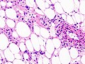
Renal AML (WC/KGH)
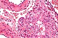
LAM - very high mag. (WC/Nephron)
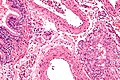
LAM - high mag. (WC/Nephron)
.jpg)


IHC
- Melanocytic markers
- Myogenic markers
- Calponin.
- Actin.[1]
- Myosin.
EM
- Premelanosomes.[4]
See also
References
- ↑ 1.0 1.1 1.2 1.3 Martignoni G, Pea M, Reghellin D, Zamboni G, Bonetti F (February 2008). "PEComas: the past, the present and the future". Virchows Arch. 452 (2): 119–32. doi:10.1007/s00428-007-0509-1. PMC 2234444. PMID 18080139. https://www.ncbi.nlm.nih.gov/pmc/articles/PMC2234444/.
- ↑ Bonetti, F.; Martignoni, G.; Colato, C.; Manfrin, E.; Gambacorta, M.; Faleri, M.; Bacchi, C.; Sin, VC. et al. (Jun 2001). "Abdominopelvic sarcoma of perivascular epithelioid cells. Report of four cases in young women, one with tuberous sclerosis.". Mod Pathol 14 (6): 563-8. doi:10.1038/modpathol.3880351. PMID 11406657.
- ↑ Doyle, LA.; Hornick, JL.; Fletcher, CD. (Dec 2013). "PEComa of the gastrointestinal tract: clinicopathologic study of 35 cases with evaluation of prognostic parameters.". Am J Surg Pathol 37 (12): 1769-82. doi:10.1097/PAS.0b013e31829caab3. PMID 24061520.
- ↑ Park, SH.; Ro, JY.; Kim, HS.; Lee, ES. (Nov 2003). "Perivascular epithelioid cell tumor of the uterus: immunohistochemical, ultrastructural and molecular study.". Pathol Int 53 (11): 800-5. PMID 14629307.