Difference between revisions of "P63"
Jump to navigation
Jump to search
(+UCC) |
|||
| Line 1: | Line 1: | ||
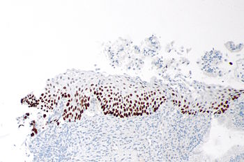
[[Image:High_grade_squamous_intraepithelial_lesion_-_2_-_p63_--_intermed_mag.jpg |thumb|right|350px| Nuclear staining is characteristic of p63.]] | |||
'''p63''' is a commonly used [[immunostain]]. p63, like most other "p" stains, is a nuclear stain. | '''p63''' is a commonly used [[immunostain]]. p63, like most other "p" stains, is a nuclear stain. | ||
Revision as of 05:24, 20 October 2014
p63 is a commonly used immunostain. p63, like most other "p" stains, is a nuclear stain.
Pattern
- Nuclear staining.
Note:
- Cytoplasmic staining suggestive of muscle differentiation - seen in rhabdomyosarcoma.[1]
Classic use
Subtype marker:
- Marker of squamous cell carcinoma.
- Urothelial carcinoma.[2]
Thresholding (invasive vs. pre-invasive):
- Prostate basal cell marker.
- Breast myoepithelial cell marker.
Non-classic tumours
- Di Como et al[3] looked at a large cross-section of tumours.
- Jo and Fletcher[4] did a paper on soft tissue lesions and p63.
References
- ↑ Martin, SE.; Temm, CJ.; Goheen, MP.; Ulbright, TM.; Hattab, EM. (Oct 2011). "Cytoplasmic p63 immunohistochemistry is a useful marker for muscle differentiation: an immunohistochemical and immunoelectron microscopic study.". Mod Pathol 24 (10): 1320-6. doi:10.1038/modpathol.2011.89. PMID 21623385.
- ↑ Lewis, JS.; Ritter, JH.; El-Mofty, S. (Nov 2005). "Alternative epithelial markers in sarcomatoid carcinomas of the head and neck, lung, and bladder-p63, MOC-31, and TTF-1.". Mod Pathol 18 (11): 1471-81. doi:10.1038/modpathol.3800451. PMID 15976812.
- ↑ Di Como, CJ.; Urist, MJ.; Babayan, I.; Drobnjak, M.; Hedvat, CV.; Teruya-Feldstein, J.; Pohar, K.; Hoos, A. et al. (Feb 2002). "p63 expression profiles in human normal and tumor tissues.". Clin Cancer Res 8 (2): 494-501. PMID 11839669.
- ↑ Jo, VY.; Fletcher, CD. (Nov 2011). "p63 immunohistochemical staining is limited in soft tissue tumors.". Am J Clin Pathol 136 (5): 762-6. doi:10.1309/AJCPXNUC7JZSKWEU. PMID 22031315.
