Osteoblastoma
Jump to navigation
Jump to search
The printable version is no longer supported and may have rendering errors. Please update your browser bookmarks and please use the default browser print function instead.
| Osteoblastoma | |
|---|---|
| Diagnosis in short | |
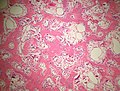
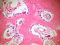
 Osteoblastoma. H&E stain. | |
|
| |
| LM | anastomosing bony trabeculae with variable mineralization, osteoblastic rimming, no nuclear atypia of osteocytes |
| LM DDx | osteoid osteoma, osteosarcoma |
| Site | bone - vertebral column typically, other bones |
|
| |
| Clinical history | usu. 15-20 years old, males > females |
| Symptoms | usu. pain |
| Radiology | often >=2.0 cm (similar lesions 1-2 cm may be osteoid osteoma), often well-circumscribed, +/-cortical expansion, +/-cortical destruction |
| Prognosis | benign, may be locally destructive |
| Clin. DDx | osteosarcoma |
Osteoblastoma is benign primary bone tumour. It is grouped with the chondro-osseous tumours.
General
- Benign bone tumour - that can be locally destructive and occasionally recurs.[1]
- Uncommon.[2]
- Typically age 15-20 and male (male:female = ~2:1).[3]
- Very large age range.[1]
- Treatment: resection.[3]
Gross
- Bone.
- Vertebral column and sacrum - most common in one large series.[1]
- Size important as per WHO definition:[4] ‡
- >= 2.0 cm: osteoblastoma.
- <=1.0 cm: osteoid osteoma.
- >1 cm and <2 cm: clinical and radiologic criteria should be considered.
Note:
- ‡1.5 cm is a diagnostic size cutoff seen in older references.[5]
Radiology
Features:
- Often well-circumscribed, +/-cortical expansion, +/-cortical destruction.[1]
Note:
- May be described as malignant by radiology.[1]
Microscopic
Features:[6]
- Anastomosing bony trabeculae with:
- Osteoblastic rimming.
- Cells line-up at edge of bone.
- Osteoblastic rimming.
Notes:
- Histomorphologically near identical/indistinguishable from osteoid osteoma.[5]
DDx:
Images
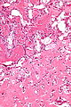
Osteoblastoma - high mag. (WC)
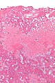
Osteoblastoma - low mag. (WC)
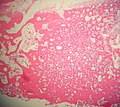
Osteoblastoma - low power. (SKB)
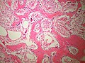
Osteoblastoma - medium power. (SKB)
Osteoblastoma - medium power. (SKB)
Osteoblastoma - Higher power - Osteoblastic rimming. (SKB)


Sign out
BONE, LEFT FEMUR, EXCISION:
- OSTEOBLASTOMA.
See also
References
- ↑ 1.0 1.1 1.2 1.3 1.4 1.5 Lucas, DR.; Unni, KK.; McLeod, RA.; O'Connor, MI.; Sim, FH. (Feb 1994). "Osteoblastoma: clinicopathologic study of 306 cases.". Hum Pathol 25 (2): 117-34. PMID 8119712.
- ↑ Khan, IS.; Thakur, JD.; Chittiboina, P.; Nanda, A.. "Large sacral osteoblastoma: a case report and review of multi-disciplinary management strategies.". J La State Med Soc 164 (5): 251-5. PMID 23362588.
- ↑ 3.0 3.1 Villalobos, CE.; Rybak, LD.; Steiner, GC.; Wittig, JC. (2010). "Osteoblastoma of the sternum--case report and review of the literature.". Bull NYU Hosp Jt Dis 68 (1): 55-9. PMID 20345366.
- ↑ Yalcinkaya, U.; Doganavsargil, B.; Sezak, M.; Kececi, B.; Argin, M.; Basdemir, G.; Oztop, F. (Dec 2014). "Clinical and morphological characteristics of osteoid osteoma and osteoblastoma: a retrospective single-center analysis of 204 patients.". Ann Diagn Pathol 18 (6): 319-25. doi:10.1016/j.anndiagpath.2014.08.006. PMID 25224389.
- ↑ 5.0 5.1 Mills, Stacey E; Carter, Darryl; Greenson, Joel K; Oberman, Harold A; Reuter, Victor E (2004). Sternberg's Diagnostic Surgical Pathology (4th ed.). Lippincott Williams & Wilkins. pp. 286. ISBN 978-0781740517.
- ↑ Mills, Stacey E; Carter, Darryl; Greenson, Joel K; Oberman, Harold A; Reuter, Victor E (2004). Sternberg's Diagnostic Surgical Pathology (4th ed.). Lippincott Williams & Wilkins. pp. 285. ISBN 978-0781740517.