Difference between revisions of "Odontogenic keratocyst"
Jump to navigation
Jump to search
(redirect) |
|||
| (4 intermediate revisions by the same user not shown) | |||
| Line 1: | Line 1: | ||
{{ Infobox diagnosis | |||
| Name = {{PAGENAME}} | |||
| Image = Keratocystic_odontogenic_tumour_-_intermed_mag.jpg | |||
| Width = | |||
| Caption = Keratocystic odontogenic tumour. [[H&E stain]]. | |||
| Synonyms = odontogenic keratocyst (old term) | |||
| Micro = stratified epithelium with "ribbon-like appearance" with palisaded basal cell layer, parakeratosis, artefactual separation of epithelium from the basement membrane | |||
| Subtypes = | |||
| LMDDx = odontogenic cyst ([[dentigerous cyst]]), [[squamous cell carcinoma]] | |||
| Stains = | |||
| IHC = | |||
| EM = | |||
| Molecular = | |||
| IF = | |||
| Gross = | |||
| Grossing = | |||
| Site = usually mandible - see ''[[odontogenic tumours and cysts]]'' | |||
| Assdx = | |||
| Syndromes = [[nevoid basal cell carcinoma syndrome]] | |||
| Clinicalhx = | |||
| Signs = mass lesion | |||
| Symptoms = | |||
| Prevalence = uncommon | |||
| Bloodwork = | |||
| Rads = | |||
| Endoscopy = | |||
| Prognosis = | |||
| Other = | |||
| ClinDDx = [[ameloblastoma]] | |||
| Tx = | |||
}} | |||
{{ Infobox external links | |||
| Name = {{PAGENAME}} | |||
| EHVSC = 10179 | |||
| EHVSC_mult = {{EHVSC2|10178}} | |||
| pathprotocols = | |||
| wikipedia = keratocystic odontogenic tumour | |||
| pathoutlines = {{Pathologyoutlines|topic/mandiblemaxillakeratocyst}} | |||
| rosaicollection = 14023 | |||
}} | |||
'''Odontogenic keratocyst''', abbreviated '''OKC''', is an uncommon [[Odontogenic tumours and cysts|odontogenic cyst]]. | |||
It was known as '''keratocystic odontogenic tumour''', abbreviated '''KOT''', from 2005 to 2017.<ref name=pmid18353202>{{Cite journal | last1 = Madras | first1 = J. | last2 = Lapointe | first2 = H. | title = Keratocystic odontogenic tumour: reclassification of the odontogenic keratocyst from cyst to tumour. | journal = J Can Dent Assoc | volume = 74 | issue = 2 | pages = 165-165h | month = Mar | year = 2008 | doi = | PMID = 18353202 }}</ref><ref name="isbn_978-92-832-2438-9">{{Citation |editor-last=El-Naggar |editor-first=Adel K |editor-last2=Chan |editor-first2=John KC |editor-last3=Grandis |editor-first3=Jennifer R |editor-last4=Takata |editor-first4=Takashi |editor-last5=Slootweg |editor-first5=Pieter J |year=2017 |title=WHO Classification of Head and Neck Tumours |edition= 4th |volume=9 |series=WHO/IARC Classification of Tumours |publisher=IARC Press |location=Lyon, France |url=http://publications.iarc.fr/Book-And-Report-Series/Who-Iarc-Classification-Of-Tumours/Who-Classification-Of-Head-And-Neck-Tumours-2017 |isbn=978-92-832-2438-9 |oclc= |lccn= |postscript=.}}</ref> | |||
==General== | |||
*May be associated with ''[[nevoid basal cell carcinoma syndrome]]''. | |||
*Very rarely transforms to a [[squamous cell carcinoma]].<ref name=pmid21493332>{{Cite journal | last1 = Lee | first1 = JW. | last2 = Gates | first2 = R. | last3 = Wignall | first3 = A. | title = Squamous cell carcinoma arising from a keratocystic odontogenic tumor. | journal = Otolaryngol Head Neck Surg | volume = 145 | issue = 2 | pages = 356-7 | month = Aug | year = 2011 | doi = 10.1177/0194599811399270 | PMID = 21493332 }}</ref><ref name=pmid23374486>{{Cite journal | last1 = Tan | first1 = B. | last2 = Yan | first2 = TS. | last3 = Shermin | first3 = L. | last4 = Teck | first4 = KC. | last5 = Yoke | first5 = PC. | last6 = Goh | first6 = C. | last7 = Balakrishnan | first7 = A. | title = Malignant transformation of keratocystic odontogenic tumor: Two case reports. | journal = Am J Otolaryngol | volume = 34 | issue = 4 | pages = 357-61 | month = | year = | doi = 10.1016/j.amjoto.2013.01.002 | PMID = 23374486 }}</ref> | |||
===Clinical=== | |||
Features:<ref name=pmid17928730>{{Cite journal | last1 = Habibi | first1 = A. | last2 = Saghravanian | first2 = N. | last3 = Habibi | first3 = M. | last4 = Mellati | first4 = E. | last5 = Habibi | first5 = M. | title = Keratocystic odontogenic tumor: a 10-year retrospective study of 83 cases in an Iranian population. | journal = J Oral Sci | volume = 49 | issue = 3 | pages = 229-35 | month = Sep | year = 2007 | doi = | PMID = 17928730 }}</ref> | |||
*Most common presentation: swelling. | |||
==Gross== | |||
*Location: usually mandible. | |||
*May mimic [[ameloblastoma]] radiologically. | |||
==Microscopic== | |||
Features: <ref>Thompson LDR. Head and neck pathology - (Foundations in diagnostic pathology). Goldblum JR, Ed.. Churchill Livingstone. 2006. ISBN 0-443-06960-3.</ref> | |||
*Stratified epithelium (resembling squamous epithelium) with: | |||
**"Ribbon-like appearance" - '''important'''. | |||
***Typically 8-10 cell layers thick - with relatively uniform thickness. | |||
***Lacks rete ridges. | |||
**Palisaded basal cell layer. | |||
*Parakeratosis (keratinized cells with nuclei) - '''key feature'''. | |||
*Artefactual separation of epithelium from the basement membrane. | |||
DDx: | |||
*Odontogenic cyst. | |||
**Orthokeratinized odontogenic cyst<ref name=pmid21062939>{{Cite journal | last1 = Macdonald-Jankowski | first1 = DS. | title = Orthokeratinized odontogenic cyst: a systematic review. | journal = Dentomaxillofac Radiol | volume = 39 | issue = 8 | pages = 455-67 | month = Dec | year = 2010 | doi = 10.1259/dmfr/19728573 | PMID = }}</ref> - usu. [[dentigerous cyst]] - has [[orthokeratosis]] instead of [[parakeratosis]]. | |||
***Orthokeratosis = keratinized cells no nuclei; parakeratosis = keratinized cell with nuclei. | |||
===Images=== | |||
<gallery> | |||
Image:Keratocystic_odontogenic_tumour_-_2_-_intermed_mag.jpg | KOT - intermed. mag. (WC) | |||
Image:Keratocystic_odontogenic_tumour_-_2_-_very_high_mag.jpg | KOT - very high mag. (WC) | |||
Image:Keratocystic_odontogenic_tumour_-_intermed_mag.jpg KOT - another case - intermed. mag. (WC)] | |||
Image:Keratocystic_odontogenic_tumour1.jpg | KOT - poor quality. (WC) | |||
Image:Keratocystic_odontogenic_tumour2.jpg | KOT - showing artefactual clefting - poor quality. (WC) | |||
</gallery> | |||
www: | |||
*[http://ars.els-cdn.com/content/image/1-s2.0-S0968605305000992-gr5.jpg KOT (els-cdn.com)].<ref>URL: [http://www.sciencedirect.com/science/article/pii/S0968605305000992#fig5 http://www.sciencedirect.com/science/article/pii/S0968605305000992#fig5]. Accessed on: 11 March 2013.</ref> | |||
==Sign out== | |||
<pre> | |||
Right Maxillary Sinus Mass, Excision: | |||
- Consistent with odontogenic keratocyst (benign ribbon-like squamous epithelium | |||
with keratinization, separated from the underlying hyaline stroma with cartilage). | |||
- Benign respiratory mucosa with mild inflammation. | |||
- NEGATIVE for malignancy. | |||
</pre> | |||
==See also== | |||
*[[Odontogenic tumours and cysts]]. | |||
*[[Head and neck pathology]]. | |||
==References== | |||
{{Reflist|2}} | |||
[[Category:Diagnosis]] | |||
[[Category:Odontogenic tumours and cysts]] | |||
Latest revision as of 21:22, 21 April 2022
| Odontogenic keratocyst | |
|---|---|
| Diagnosis in short | |
 Keratocystic odontogenic tumour. H&E stain. | |
|
| |
| Synonyms | odontogenic keratocyst (old term) |
|
| |
| LM | stratified epithelium with "ribbon-like appearance" with palisaded basal cell layer, parakeratosis, artefactual separation of epithelium from the basement membrane |
| LM DDx | odontogenic cyst (dentigerous cyst), squamous cell carcinoma |
| Site | usually mandible - see odontogenic tumours and cysts |
|
| |
| Syndromes | nevoid basal cell carcinoma syndrome |
|
| |
| Signs | mass lesion |
| Prevalence | uncommon |
| Clin. DDx | ameloblastoma |
| Odontogenic keratocyst | |
|---|---|
| External resources | |
| EHVSC | 10179 10178 |
| Wikipedia | keratocystic odontogenic tumour |
| Pathology Outlines | topic/mandiblemaxillakeratocyst |
| Rosai Collection | 14023 |
Odontogenic keratocyst, abbreviated OKC, is an uncommon odontogenic cyst.
It was known as keratocystic odontogenic tumour, abbreviated KOT, from 2005 to 2017.[1][2]
General
- May be associated with nevoid basal cell carcinoma syndrome.
- Very rarely transforms to a squamous cell carcinoma.[3][4]
Clinical
Features:[5]
- Most common presentation: swelling.
Gross
- Location: usually mandible.
- May mimic ameloblastoma radiologically.
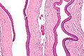
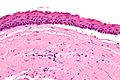
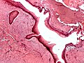
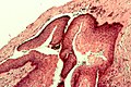
Microscopic
Features: [6]
- Stratified epithelium (resembling squamous epithelium) with:
- "Ribbon-like appearance" - important.
- Typically 8-10 cell layers thick - with relatively uniform thickness.
- Lacks rete ridges.
- Palisaded basal cell layer.
- "Ribbon-like appearance" - important.
- Parakeratosis (keratinized cells with nuclei) - key feature.
- Artefactual separation of epithelium from the basement membrane.
DDx:
- Odontogenic cyst.
- Orthokeratinized odontogenic cyst[7] - usu. dentigerous cyst - has orthokeratosis instead of parakeratosis.
- Orthokeratosis = keratinized cells no nuclei; parakeratosis = keratinized cell with nuclei.
- Orthokeratinized odontogenic cyst[7] - usu. dentigerous cyst - has orthokeratosis instead of parakeratosis.
Images
KOT - intermed. mag. (WC)
KOT - very high mag. (WC)
KOT - poor quality. (WC)
KOT - showing artefactual clefting - poor quality. (WC)


www:
Sign out
Right Maxillary Sinus Mass, Excision:
     - Consistent with odontogenic keratocyst (benign ribbon-like squamous epithelium
       with keratinization, separated from the underlying hyaline stroma with cartilage).
     - Benign respiratory mucosa with mild inflammation.
     - NEGATIVE for malignancy.
See also
References
- ↑ Madras, J.; Lapointe, H. (Mar 2008). "Keratocystic odontogenic tumour: reclassification of the odontogenic keratocyst from cyst to tumour.". J Can Dent Assoc 74 (2): 165-165h. PMID 18353202.
- ↑ El-Naggar, Adel K, ed. (2017), WHO Classification of Head and Neck Tumours, WHO/IARC Classification of Tumours, 9 (4th ed.), Lyon, France: IARC Press, ISBN 978-92-832-2438-9, http://publications.iarc.fr/Book-And-Report-Series/Who-Iarc-Classification-Of-Tumours/Who-Classification-Of-Head-And-Neck-Tumours-2017.
- ↑ Lee, JW.; Gates, R.; Wignall, A. (Aug 2011). "Squamous cell carcinoma arising from a keratocystic odontogenic tumor.". Otolaryngol Head Neck Surg 145 (2): 356-7. doi:10.1177/0194599811399270. PMID 21493332.
- ↑ Tan, B.; Yan, TS.; Shermin, L.; Teck, KC.; Yoke, PC.; Goh, C.; Balakrishnan, A.. "Malignant transformation of keratocystic odontogenic tumor: Two case reports.". Am J Otolaryngol 34 (4): 357-61. doi:10.1016/j.amjoto.2013.01.002. PMID 23374486.
- ↑ Habibi, A.; Saghravanian, N.; Habibi, M.; Mellati, E.; Habibi, M. (Sep 2007). "Keratocystic odontogenic tumor: a 10-year retrospective study of 83 cases in an Iranian population.". J Oral Sci 49 (3): 229-35. PMID 17928730.
- ↑ Thompson LDR. Head and neck pathology - (Foundations in diagnostic pathology). Goldblum JR, Ed.. Churchill Livingstone. 2006. ISBN 0-443-06960-3.
- ↑ Macdonald-Jankowski, DS. (Dec 2010). "Orthokeratinized odontogenic cyst: a systematic review.". Dentomaxillofac Radiol 39 (8): 455-67. doi:10.1259/dmfr/19728573.
- ↑ URL: http://www.sciencedirect.com/science/article/pii/S0968605305000992#fig5. Accessed on: 11 March 2013.