Difference between revisions of "Neurothekeoma"
Jump to navigation
Jump to search
(redirect) |
|||
| (11 intermediate revisions by the same user not shown) | |||
| Line 1: | Line 1: | ||
'''Neurothekeoma''' is a benign [[peripheral nerve sheath tumours|peripheral nerve sheath tumour]]. | |||
It is also known as '''nerve sheath myxoma'''<ref name=pmid11070116>{{Cite journal | last1 = Laskin | first1 = WB. | last2 = Fetsch | first2 = JF. | last3 = Miettinen | first3 = M. | title = The "neurothekeoma": immunohistochemical analysis distinguishes the true nerve sheath myxoma from its mimics. | journal = Hum Pathol | volume = 31 | issue = 10 | pages = 1230-41 | month = Oct | year = 2000 | doi = 10.1053/hupa.2000.18474 | PMID = 11070116 }}</ref> and '''myxoma of the nerve sheath'''. | |||
There is growing evidence that neurothekomas and dermal nerve sheath myxomas are distinct entities.<ref>{{Cite journal | last1 = Sheth | first1 = S. | last2 = Li | first2 = X. | last3 = Binder | first3 = S. | last4 = Dry | first4 = SM. | title = Differential gene expression profiles of neurothekeomas and nerve sheath myxomas by microarray analysis. | journal = Mod Pathol | volume = 24 | issue = 3 | pages = 343-54 | month = Mar | year = 2011 | doi = 10.1038/modpathol.2010.203 | PMID = 21297585 }}</ref> | |||
==General== | |||
*Rare. | |||
*Female > male. | |||
==Microscopic== | |||
Features:<ref name=pmid17325474>{{cite journal |author=Hornick JL, Fletcher CD |title=Cellular neurothekeoma: detailed characterization in a series of 133 cases |journal=Am. J. Surg. Pathol. |volume=31 |issue=3 |pages=329–40 |year=2007 |month=March |pmid=17325474 |doi=10.1097/01.pas.0000213360.03133.89 |url=}}</ref> | |||
*Superficial dermal lesion: | |||
**Usu. lobulated or micronodular architecture - '''key feature'''. | |||
***+/-Focal sheeting. | |||
**Spindle/epithelioid morphology with pale eosinophilic cytoplasm - '''key feature'''. | |||
**+/-Inflammation around lesion. | |||
**+/-Surrounded by collagen. | |||
Notes: | |||
*No atypia. | |||
*Mitoses rare/none. | |||
*Often poorly circumscribed. | |||
Subtypes:<ref name=pmid10555009>{{cite journal |author=Wang AR, May D, Bourne P, Scott G |title=PGP9.5: a marker for cellular neurothekeoma |journal=Am. J. Surg. Pathol. |volume=23 |issue=11 |pages=1401–7 |year=1999 |month=November |pmid=10555009 |doi= |url=}}</ref> | |||
*Cellular. | |||
*[[Myxoid]]. | |||
*Intermediate. | |||
DDx: | |||
*[[Dermatofibroma]]. | |||
*[[Angiomatoid fibrous histiocytoma]] -- have cystic blood filled spaces, inflammation.<ref>URL: [http://surgpathcriteria.stanford.edu/softfib/angiomatoid_fibrous_histiocytoma/ http://surgpathcriteria.stanford.edu/softfib/angiomatoid_fibrous_histiocytoma/]. Accessed on: 11 May 2011.</ref> | |||
===Images=== | |||
<gallery> | |||
File:Neurothekeoma2.JPG | Neurothekeoma (WP) | |||
</gallery> | |||
====www==== | |||
*[http://path.upmc.edu/cases/case586.html Neurothekeoma - several images (upmc.edu)]. | |||
*[http://www.dermpedia.org/files/images/Image2_49.jpg Nerve sheath myxoma (dermpedia)]] | |||
==IHC== | |||
Features:<ref name=pmid17325474/> | |||
*NKI/C3 ([[AKA]] NKI-C3) +ve. | |||
*NSE +/-ve. | |||
Others:<ref name=pmid17592278>{{cite journal |author=Fetsch JF, Laskin WB, Hallman JR, Lupton GP, Miettinen M |title=Neurothekeoma: an analysis of 178 tumors with detailed immunohistochemical data and long-term patient follow-up information |journal=Am. J. Surg. Pathol. |volume=31 |issue=7 |pages=1103–14 |year=2007 |month=July |pmid=17592278 |doi=10.1097/PAS.0b013e31802d96af |url=}}</ref> | |||
*Vimentin +ve. | |||
*CD10 +ve. | |||
*[[Microphthalmia transcription factor]] (MITF) +ve. | |||
*PGP9.5 +ve. | |||
*S100 +ve.<ref name=RefDerm541>{{Ref Derm|541}}</ref> | |||
**-ve in older literature. | |||
==See also== | |||
*[[Peripheral nerve sheath tumours]]. | |||
==References== | |||
{{Reflist|1}} | |||
[[Category:Diagnosis]] | |||
Latest revision as of 15:29, 5 November 2021
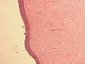
Neurothekeoma is a benign peripheral nerve sheath tumour.
It is also known as nerve sheath myxoma[1] and myxoma of the nerve sheath.
There is growing evidence that neurothekomas and dermal nerve sheath myxomas are distinct entities.[2]
General
- Rare.
- Female > male.
Microscopic
Features:[3]
- Superficial dermal lesion:
- Usu. lobulated or micronodular architecture - key feature.
- +/-Focal sheeting.
- Spindle/epithelioid morphology with pale eosinophilic cytoplasm - key feature.
- +/-Inflammation around lesion.
- +/-Surrounded by collagen.
- Usu. lobulated or micronodular architecture - key feature.
Notes:
- No atypia.
- Mitoses rare/none.
- Often poorly circumscribed.
Subtypes:[4]
- Cellular.
- Myxoid.
- Intermediate.
DDx:
- Dermatofibroma.
- Angiomatoid fibrous histiocytoma -- have cystic blood filled spaces, inflammation.[5]
Images
Neurothekeoma (WP)

www
IHC
Features:[3]
- NKI/C3 (AKA NKI-C3) +ve.
- NSE +/-ve.
Others:[6]
- Vimentin +ve.
- CD10 +ve.
- Microphthalmia transcription factor (MITF) +ve.
- PGP9.5 +ve.
- S100 +ve.[7]
- -ve in older literature.
See also
References
- ↑ Laskin, WB.; Fetsch, JF.; Miettinen, M. (Oct 2000). "The "neurothekeoma": immunohistochemical analysis distinguishes the true nerve sheath myxoma from its mimics.". Hum Pathol 31 (10): 1230-41. doi:10.1053/hupa.2000.18474. PMID 11070116.
- ↑ Sheth, S.; Li, X.; Binder, S.; Dry, SM. (Mar 2011). "Differential gene expression profiles of neurothekeomas and nerve sheath myxomas by microarray analysis.". Mod Pathol 24 (3): 343-54. doi:10.1038/modpathol.2010.203. PMID 21297585.
- ↑ 3.0 3.1 Hornick JL, Fletcher CD (March 2007). "Cellular neurothekeoma: detailed characterization in a series of 133 cases". Am. J. Surg. Pathol. 31 (3): 329–40. doi:10.1097/01.pas.0000213360.03133.89. PMID 17325474.
- ↑ Wang AR, May D, Bourne P, Scott G (November 1999). "PGP9.5: a marker for cellular neurothekeoma". Am. J. Surg. Pathol. 23 (11): 1401–7. PMID 10555009.
- ↑ URL: http://surgpathcriteria.stanford.edu/softfib/angiomatoid_fibrous_histiocytoma/. Accessed on: 11 May 2011.
- ↑ Fetsch JF, Laskin WB, Hallman JR, Lupton GP, Miettinen M (July 2007). "Neurothekeoma: an analysis of 178 tumors with detailed immunohistochemical data and long-term patient follow-up information". Am. J. Surg. Pathol. 31 (7): 1103–14. doi:10.1097/PAS.0b013e31802d96af. PMID 17592278.
- ↑ Busam, Klaus J. (2009). Dermatopathology: A Volume in the Foundations in Diagnostic Pathology Series (1st ed.). Saunders. pp. 541. ISBN 978-0443066542.