Neurohistology
Jump to navigation
Jump to search
This article covers basic (normal) neurohistology. It is essential to have a good grasp on neurohistology... before doing neuropathology.
Normal cells
- Neuron:
- Abundant cytoplasm - key feature.
- Often very large cells, with angled edges.
- Prominent nucleolus.
- Nissl substance (granular perinuclear material - rough ER).
- Glial cells.
- Oligodendrocyte.
- Small round nuclei (lymphocyte-like nucleus) - key feature.
- May resemble a fried egg on H&E (clear cytoplasm, central nucleus).
- Astrocyte.
- Irregular non-ovoid nucleus - key feature.
- Nuclei less dense than in oligodendrocyte.
- Close to blood vessels.
- Form blood-brain barrier.
- Cytoplasm normally not visible.
- Image: astrocyte (med.unsw.edu.au) (in endocrine development).
- Microglia - macrophage of the brain (derived from monocyte).
- May be large.
- May have vesicles.
- Rarely seen in normal tissue.
- Oligodendrocyte.
- Ependyma.
- Simple ciliated cuboidal epithelium.
- Image: Ependyma (stonybrookmedicalcenter.org).
Normal cellular constituents in a table
| Key feature | Other features | Image | |
| Neuron | cytoplasm | Nissl substance (prominent RER), "sharp" corners in cell membrane, nucleolus - usu. prominent[1] |
red neurons (WC) |
| Astrocyte | non-ovoid nucleus | no cytoplasm | (unsw.edu) |
| Oligodendrocyte | round small nucleus | peri-nuclear clearing | |
| Microglia | rod-like shape, may have "bent" nucleus |
rarely seen in normal tissue | (ucsf.edu),(vcu.edu) |
Neurons
There are many types of 'em. Broadly, they can be classified as:
- Pyramidal - have a pyramidal shape.
- Dentrites go to molecular layer.
- Axons go to outside of cortex.
- Non-pyramidal.
Motor neurons:
- Coarse Nissl substance - key feature.
- Nissl described as having a tigroid appearance.[2]
- Polygonal shape.
- Send dendrites in all directions.
Histology by anatomical structure
Subependyma
Features:[3]
- Ependyma (simple ciliated cuboidal epithelium).
- Subependymal plate - connective tissue with blood vessels.
Pons
Features:
- Looks like bacon.[4]
- Image: Pons (stonybrookmedicalcenter.org).
Caudate
Features:
- Neurons with adjacent ependymal lining.[5]
- The caudate forms lateral wall of lateral ventricle.
Putamen
Features:
- Histologically identical to the caudate - but not adjacent to a ventricle, i.e. an ependymal lining.
- Striatopallidal fibers AKA pencils of Wilson - bundles of blue fibres (on H&E LFB).
Globus pallidus
Features:
- Histologically distinct from caudate and putamen.
Hippocampus
Hippocampal formation:[6]
- Dentate gyrus.
- "Dense" thin layer of nuclei.
- Quasi "U-shaped"; "open" (top) portion of "U" is superolateral.
- Image: Dentate gyrus (stonybrookmedicalcenter.org).
- Hippocampus proper (AKA Ammon's horn) - this is subdivided:
- CA3 - superior.
- CA1 - inferior (next to subiculum).
- CA2 - in between CA3 and CA1, lateral.
- CA4 - medial (closest to dentate gyrus; CA4 sits in "open" part of "U").
- Subicular complex.
Images:
- Hippocampus - frontal section (WP).
- Hippocampus - good schematic (WC).
- Hippocampus (ajnr.org).
- Hippocampus and subiculum (hu-berlin.de).
- Hippocampus - crappy schematic (ucsd.edu).
Important note:
- CA1 - weak link, dies in ischemia, affected by hypoglycemia.
- CA2 - resistant to ischemia.
DDx of ischemia-like changes in the hippocampus:
- Toxins.
Cerebellum
Main components:
- Cortex (superficial) - branches (Christmas tree-like).
- Dentate nucleus (deep) - looks like the bite impression of a molar.
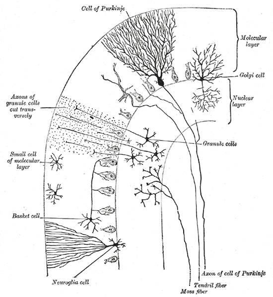
Cerebellar cortex:
- Layers (superficial to deep) - mnemonic MPG:[7]
- Molecular layer -- "very pink" on H&E.
- Inhibitory interneurons: stellate cells, basket cells.
- Purkinje cell layer.
- One cell layer thick - hueuege cells (~50-80 micrometers[1]).
- Very large nucleus (~4x RBC diameter =~ 4x the size of granule cell).
- Large nucleolus (~1x RBC diameter =~ size of granule cell).
- Very large nucleus (~4x RBC diameter =~ 4x the size of granule cell).
- One cell layer thick - hueuege cells (~50-80 micrometers[1]).
- Granule cell layer -- "very blue" on H&E.
- Granule cells (many), interneurons (Golgi cells --few in number). (???)
- Molecular layer -- "very pink" on H&E.
- Images:
Notes:
- Bergmann glia are found between the molecular layer & granular layer. They are normally not seen. They are increased & prominent in pathologic states (e.g. ischemia); "Bergmann gliosis".[8]
Cerebral cortex
Layers (superficial to deep):
- Molecular layer.
- Empty appearing.
- Outer granular layer.
- Higher cell density & smaller cells than pyramidal layer.
- Outer pyramidal layer.
- Inner granular layer.
- Not prominent in frontal cortex.
- Where the thalamic axons end.
- Divided in three (a, b, c) in the calcarine cortex due to two white matter bands (external band of Baillarger, internal band of Baillarger) than are grossly identified as the line of Gennari.[9]
- Inner pyramidal layer.
- Location of Betz neurons - large motor neurons of cerebral cortex.
- Multiforme layer (Polymorphic layer).
Images:
See also
References
- ↑ 1.0 1.1 Perry, Arie; Brat, Daniel J. (2010). Practical Surgical Neuropathology: A Diagnostic Approach: A Volume in the Pattern Recognition series (1st ed.). Churchill Livingstone. pp. 16. ISBN 978-0443069826.
Cite error: Invalid
<ref>tag; name "Ref_PSNP16" defined multiple times with different content - ↑ URL: http://www.stonybrookmedicalcenter.org/pathology/neuropathology/chapter1. Accessed on: 5 July 2010.
- ↑ Half-day. 28 June 2010.
- ↑ Half-day. 28 June 2010.
- ↑ URL: http://www.stonybrookmedicalcenter.org/pathology/neuropathology/chapter1. Accessed on: 2 July 2010.
- ↑ URL: http://www.stonybrookmedicalcenter.org/pathology/neuropathology/chapter1. Accessed on: 2 July 2010.
- ↑ URL: http://www.stonybrookmedicalcenter.org/pathology/neuropathology/chapter1. Accessed on: 2 July 2010.
- ↑ Perry, Arie; Brat, Daniel J. (2010). Practical Surgical Neuropathology: A Diagnostic Approach: A Volume in the Pattern Recognition series (1st ed.). Churchill Livingstone. pp. 18. ISBN 978-0443069826.
- ↑ Perry, Arie; Brat, Daniel J. (2010). Practical Surgical Neuropathology: A Diagnostic Approach: A Volume in the Pattern Recognition series (1st ed.). Churchill Livingstone. pp. 24. ISBN 978-0443069826.