Difference between revisions of "Neurohistology"
(→Locus ceruleus: more) |
Jensflorian (talk | contribs) (→Glial cells: microglia) |
||
| (35 intermediate revisions by 2 users not shown) | |||
| Line 7: | Line 7: | ||
==Overview== | ==Overview== | ||
===Central nervous system=== | |||
====Neuron==== | |||
*Abundant cytoplasm - '''key feature'''. | |||
*Often very large cells, with angled edges. | |||
*Prominent nucleolus. | |||
*Nissl substance (granular perinuclear material = rough endoplasmic reticulum). | |||
====Glial cells==== | |||
*Oligodendrocyte. | |||
**Astrocyte. | **Small round nuclei (lymphocyte-like nucleus) - '''key feature'''. | ||
**May resemble a ''fried egg'' on H&E (clear cytoplasm, central nucleus). | |||
**Image: [http://www.urmc.rochester.edu/libraries/courses/neuroslides/lab1a/images/1-11.png oligodendrocyte urmc.rochester.edu] | |||
*Astrocyte. | |||
**Irregular non-ovoid nucleus - '''key feature'''. | |||
**Nuclei less dense than in oligodendrocyte. | |||
**Close to blood vessels. | |||
**Form blood-brain barrier. | |||
** | **Cytoplasm normally ''not'' visible. | ||
*** | **Image: [http://embryology.med.unsw.edu.au/histology/endocrine/pin42he.jpg astrocyte (med.unsw.edu.au)] (in [http://embryology.med.unsw.edu.au/notes/endocrine12.htm endocrine development]). | ||
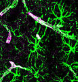
*Microglia - macrophage of the brain (derived from monocyte). | |||
**Typically large cells with abundant cytoplasm. | |||
***Often have vesicles. | |||
**Rarely seen in normal tissue. | |||
**Three morphologic types: | |||
***Ramified microglia. | |||
***Hypertrophic microglia. | |||
***Dystrophic microglia - often seen in neurodegnerative disease. | |||
*Ependyma. | *Ependyma. | ||
**Simple ciliated cuboidal epithelium. | **Simple ciliated cuboidal epithelium. | ||
**Image: [http://www.stonybrookmedicalcenter.org/sbumcfiles/images/221-001.jpg Ependyma (stonybrookmedicalcenter.org)]. | **Image: [http://www.stonybrookmedicalcenter.org/sbumcfiles/images/221-001.jpg Ependyma (stonybrookmedicalcenter.org)]. | ||
*Choroid plexus. | |||
**Specialized ependymal cells | |||
**Cuboidal epithelial cells surrounding a core of capillaries and loose connective tissue. | |||
**Ventricular location. | |||
=====Images===== | |||
[[File:Blausen 0870 TypesofNeuroglia.png |500px| Types of neuroglia (WC/Blausen)]] | |||
<gallery> | |||
File:Astrocytes.jpg | Astrocytes highlighted by GFAP (WC/Jeffery J. Iliff) | |||
Image:Oligodendrocyte_HE_stain_high_mag.jpg | Oligodendrocytes with fried-egg appearance (WC/jensflorian) | |||
Image:Ependyma.png|Ependymal cells. (WC/marvin101) | |||
File:Psammoma bodies in the choroid plexus, HE.JPG | Choroid plexus (WC/Patho) | |||
File:Mikroglej 1.jpg | Microglial cells, Lectin stain (WC/Grzegorz Wicher) | |||
</gallery> | |||
===Peripheral nervous system=== | |||
====Ganglion cell==== | |||
*Nerve cell body. | |||
*Found in ganglions - encapsulated body ~ 100-300 μm. | |||
*Large round nucleus with prominent nucleolus - '''key feature'''. | |||
*Abundant eosinophilic granular cytoplasm. | |||
====Schwann cell==== | |||
*Principal glia of the PNS. | |||
*Myelinated or unmyelinated. | |||
*Ovoid, wavy nuclei. | |||
*Cells have a basal membrane. | |||
*Found in peripheral nerves. | |||
*Aka ''neurolemmocytes''. | |||
=====Images===== | |||
<gallery> | |||
Image:Ganglion_very_high_mag.jpg | Ganglion - very high mag. (WC/Nephron) | |||
Image:Peripheral nerve, cross section.jpg | Peripheral nerve - cross section. (WC/Reytan) | |||
File:Nerve root - very high mag.jpg | Nerve root HPS stain (WC/Nephron) | |||
</gallery> | |||
WC: | |||
*[http://commons.wikimedia.org/wiki/File:Ganglion_intermed_mag.jpg Ganglion - intermed. mag. (WC/Nephron)]. | |||
==Normal cellular constituents in a table== | ==Normal cellular constituents in a table== | ||
{| class="wikitable" | {| class="wikitable sortable" | ||
! Cell | |||
! Key feature | |||
! Other features | |||
! Image | |||
|- | |- | ||
|Neuron | |Neuron | ||
| Line 73: | Line 116: | ||
*Send dendrites in all directions. | *Send dendrites in all directions. | ||
Images: | ===Images=== | ||
www: | |||
*[http://www.stonybrookmedicalcenter.org/sbumcfiles/images/238-002.jpg Motor neuron (stonybrookmedicalcenter.org)]. | *[http://www.stonybrookmedicalcenter.org/sbumcfiles/images/238-002.jpg Motor neuron (stonybrookmedicalcenter.org)]. | ||
<gallery> | |||
Image:Medulla_oblongata_-_posterior_-_cn_xii_-_very_high_mag.jpg | Motor neurons - nucleus of CN XII - very high mag. (WC) | |||
</gallery> | |||
=Structures= | =Structures= | ||
This section deals with structures seen at several places in the CNS. | This section deals with structures seen at several places in the CNS. | ||
==Grey | ==Grey matter and white matter== | ||
{| class="wikitable" | {| class="wikitable" | ||
| | | | ||
| Line 95: | Line 141: | ||
|- | |- | ||
| Image | | Image | ||
| [ | | [[Image:Grey_matter_and_white_matter_-_very_high_mag.jpg|thumb|Right 2/3 of image. (WC/Nephron)]] | ||
| [ | | [[Image:Grey_matter_and_white_matter_-_very_high_mag.jpg|thumb|Left 1/3 of image. (WC/Nephron)]] | ||
|} | |} | ||
Additional images (white matter vs. grey matter): | Additional images (white matter vs. grey matter): | ||
<gallery> | |||
Image:Grey_matter_and_white_matter_-_high_mag.jpg | Grey-white matter interface - high mag. (WC) | |||
Image:Grey_matter_and_white_matter_-_intermed_mag.jpg | Grey and white matter - intermed. mag. (WC) | |||
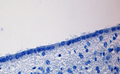
File:LFB_CNS_cortex_supratentorial.jpg | Normal cortex - LFB. (WC/jensflorian) | |||
File:LFB_CNS_cortex_grey-white_matter_junction.jpg | White-grey matter junction - LFB. (WC/jensflorian) | |||
</gallery> | |||
==Vessels== | ==Vessels== | ||
| Line 127: | Line 177: | ||
*#Small - GABA. | *#Small - GABA. | ||
*#Large (very rare: ~1 in 100-200) - cholingeric. | *#Large (very rare: ~1 in 100-200) - cholingeric. | ||
Notes: | Notes: | ||
*Histologically identical to the ''caudate'' - but not adjacent to a ventricle. | *Histologically identical to the ''caudate'' - but not adjacent to a ventricle. | ||
**The caudate is adjacent to an ependymal lined space, putamen is not.<ref>URL: [http://www.stonybrookmedicalcenter.org/pathology/neuropathology/chapter1 http://www.stonybrookmedicalcenter.org/pathology/neuropathology/chapter1]. Accessed on: 22 December 2010.</ref> | **The caudate is adjacent to an ependymal lined space, putamen is not.<ref>URL: [http://www.stonybrookmedicalcenter.org/pathology/neuropathology/chapter1 http://www.stonybrookmedicalcenter.org/pathology/neuropathology/chapter1]. Accessed on: 22 December 2010.</ref> | ||
*[[Necrotic]] putamen in methanol poisoning.<ref name=pmid19015300>{{Cite journal | last1 = Bhatia | first1 = R. | last2 = Kumar | first2 = M. | last3 = Garg | first3 = A. | last4 = Nanda | first4 = A. | title = Putaminal necrosis due to methanol toxicity. | journal = Pract Neurol | volume = 8 | issue = 6 | pages = 386-7 | month = Dec | year = 2008 | doi = 10.1136/jnnp.2008.161976 | PMID = 19015300 }}</ref> | |||
===Images=== | |||
<gallery> | |||
Image:Globus_pallidus_and_putamen_-_very_low_mag.jpg | Globus pallidus & putamen - showing Wilson's pencils - very low mag. (WC) | |||
Image:Putamen_-_intermed_mag.jpg | Putamen - intermed. mag. (WC) | |||
</gallery> | |||
www: | |||
*[http://frontalcortex.com/?page=oll&topic=24&qid=760 Pencils of Wilson (frontalcortex.com)]. | |||
==Globus pallidus== | ==Globus pallidus== | ||
| Line 147: | Line 200: | ||
*Histologically distinct from caudate and putamen. | *Histologically distinct from caudate and putamen. | ||
Image | ===Image=== | ||
<gallery> | |||
Image:Nucleus_basalis_of_Meynert_-l-_very_low_mag.jpg | Globus pallidus, putamen and the nucleus basalis in the substantia innominata - very low mag. (WC) | |||
</gallery> | |||
==Hippocampus== | ==Hippocampus== | ||
| Line 167: | Line 223: | ||
#*Transitions to the six layers in the ''entorhinal cortex''. | #*Transitions to the six layers in the ''entorhinal cortex''. | ||
#**No vacuolated looking stuff next to it. | #**No vacuolated looking stuff next to it. | ||
Important notes: | Important notes: | ||
| Line 179: | Line 228: | ||
*CA2 - resistant to ischemia. | *CA2 - resistant to ischemia. | ||
*CA4 - involved in [[epilepsy]],<ref name=pmid3518570>{{Cite journal | last1 = Ingvar | first1 = M. | title = Cerebral blood flow and metabolic rate during seizures. Relationship to epileptic brain damage. | journal = Ann N Y Acad Sci | volume = 462 | issue = | pages = 194-206 | month = | year = 1986 | doi = | PMID = 3518570 }}</ref> usu. normal in degenerative diseases.<ref>D. Munoz. 27 July 2011.</ref> | *CA4 - involved in [[epilepsy]],<ref name=pmid3518570>{{Cite journal | last1 = Ingvar | first1 = M. | title = Cerebral blood flow and metabolic rate during seizures. Relationship to epileptic brain damage. | journal = Ann N Y Acad Sci | volume = 462 | issue = | pages = 194-206 | month = | year = 1986 | doi = | PMID = 3518570 }}</ref> usu. normal in degenerative diseases.<ref>D. Munoz. 27 July 2011.</ref> | ||
====Images==== | |||
<gallery> | |||
Image:Brainmaps-macaque-hippocampus.jpg | Hippocampus - frontal section. (WC) | |||
Image:Hippocampus_%28brain%29.jpg | Hippocampus - schematic. (WC) | |||
</gallery> | |||
www: | |||
*[http://www.ajnr.org/cgi/content-nw/full/28/5/958/F1 Hippocampus (ajnr.org)]. | |||
*[http://edoc.hu-berlin.de/dissertationen/schubert-stephan-nicolas-2003-09-26/HTML/schubert_html_21d219b1.png Hippocampus and subiculum (hu-berlin.de)]. | |||
*[http://spinwarp.ucsd.edu/NeuroWeb/Text/br-800epi/br-800epi1.gif Hippocampus - crappy schematic (ucsd.edu)]. | |||
===Layers of CA<ref name=Ref_PSNP25>{{Ref PSNP|25}}</ref>=== | ===Layers of CA<ref name=Ref_PSNP25>{{Ref PSNP|25}}</ref>=== | ||
| Line 198: | Line 256: | ||
**Small neurons. | **Small neurons. | ||
Images | ====Images==== | ||
<gallery> | |||
Image:Dentate_nucleus_-_cerebellum_-_intermed_mag.jpg | Dentate nucleus of the cerebellum - intermed. mag. (WC) | |||
Image:Dentate_nucleus_-_cerebellum_-_high_mag.jpg | Dentate nucleus of the cerebellum - high mag. (WC) | |||
</gallery> | |||
===Cerebellar cortex=== | ===Cerebellar cortex=== | ||
| Line 214: | Line 274: | ||
*#**May look like small cell carcinoma to the uninitiated. | *#**May look like small cell carcinoma to the uninitiated. | ||
*#*Golgi cells (interneurons) - few in number, elongated/columnar, 3-5x size of granule cell. | *#*Golgi cells (interneurons) - few in number, elongated/columnar, 3-5x size of granule cell. | ||
Notes: | Notes: | ||
*''Bergmann glia'' are found between the molecular layer & granular layer. They are normally not seen. They are increased & prominent in pathologic states (e.g. ischemia); "[[Bergmann gliosis]]".<ref name=Ref_PSNP18>{{Ref PSNP|18}}</ref> | *''Bergmann glia'' are found between the molecular layer & granular layer. They are normally not seen. They are increased & prominent in pathologic states (e.g. ischemia); "[[Bergmann gliosis]]".<ref name=Ref_PSNP18>{{Ref PSNP|18}}</ref> | ||
====Images==== | |||
www: | |||
*[http://www.stonybrookmedicalcenter.org/sbumcfiles/images/227_001.jpg Cerebellar cortex - micrograph with labels (stonybrookmedicalcenter.org)]. | |||
<gallery> | |||
Image:Gray706.png | Cerebellar cortex - schematic. (Gray's Anatomy/WC) | |||
Image:Cerebellar_cortex_-_intermed_mag.jpg | Cerebellar cortex - intermed. mag. (WC) | |||
Image:Cerebellum_-_biel_-_high_mag.jpg | Cerebellar cortex - high mag. [[Bielschowsky stain]]. (WC) | |||
</gallery> | |||
==Cerebral cortex== | ==Cerebral cortex== | ||
| Line 246: | Line 310: | ||
*[http://williamcalvin.com/bk7/img/bk7p31.jpg Cerebral cortex (williamcalvin.com)]. | *[http://williamcalvin.com/bk7/img/bk7p31.jpg Cerebral cortex (williamcalvin.com)]. | ||
*[http://www.benbest.com/science/anatmind/anatmd5.html Cerebral cortex (benbest.com)]. | *[http://www.benbest.com/science/anatmind/anatmd5.html Cerebral cortex (benbest.com)]. | ||
===Cingulate cortex=== | |||
*Spindle neurons, [[AKA]] ''von Economo neurons''. | |||
**Thought to be important in cognition and problem solving.<ref name=pmid11411161>{{Cite journal | last1 = Allman | first1 = JM. | last2 = Hakeem | first2 = A. | last3 = Erwin | first3 = JM. | last4 = Nimchinsky | first4 = E. | last5 = Hof | first5 = P. | title = The anterior cingulate cortex. The evolution of an interface between emotion and cognition. | journal = Ann N Y Acad Sci | volume = 935 | issue = | pages = 107-17 | month = May | year = 2001 | doi = | PMID = 11411161 }}</ref> | |||
====Images==== | |||
<gallery> | |||
Image:Spindle_neurons_-_high_mag.jpg | Spindle neurons - high mag. (WC) | |||
Image:Spindle_neurons_-_very_high_mag_-_cropped.jpg | Spindle neurons - cropped - very high mag. (WC) | |||
</gallery> | |||
==Pineal gland== | ==Pineal gland== | ||
| Line 256: | Line 330: | ||
*Astrocytes: | *Astrocytes: | ||
**Cylindrical hyperchromatic nucleus ~ 1/2 the size of pinealocyte. | **Cylindrical hyperchromatic nucleus ~ 1/2 the size of pinealocyte. | ||
Notes: | Notes: | ||
*Highly cellular structure - may be confused with (metastatic) small cell carcinoma. | *Highly cellular structure - may be confused with (metastatic) [[small cell carcinoma]]. | ||
*Often calcified. | *Often calcified. | ||
===Images=== | |||
<gallery> | |||
Image:Pineal_gland_-_very_high_mag.jpg | Pineal gland - very high mag. (WC/Nephron) | |||
Image:Pineal_gland_-_intermed_mag.jpg | Pineal gland - intermed. mag. (WC/Nephron) | |||
Image:Pineal.jpg | Pineal gland. (WC) | |||
</gallery> | |||
www: | |||
*[http://www.lab.anhb.uwa.edu.au/mb140/corepages/endocrines/Images/pin42he.jpg Pineal gland (anhb.uwa.edu.au)].<ref name=Blue_Histology_-_Endocrines>URL: [http://www.lab.anhb.uwa.edu.au/mb140/corepages/endocrines/endocrin.htm http://www.lab.anhb.uwa.edu.au/mb140/corepages/endocrines/endocrin.htm]. Accessed on: 31 October 2010.</ref> | |||
===IHC=== | ===IHC=== | ||
*Synaptophysin +ve.<ref name=Ref_PSNP25-26>{{Ref_PSNP|25-26}}</ref> | *Synaptophysin +ve.<ref name=Ref_PSNP25-26>{{Ref_PSNP|25-26}}</ref> | ||
| Line 272: | Line 347: | ||
==Midbrain== | ==Midbrain== | ||
Structures: | Structures: | ||
*Substantia nigra (Parkinson's disease). | *Substantia nigra ([[Parkinson's disease]]). | ||
*Nuclei of CN IV (posterior). | *Nuclei of CN IV (posterior). | ||
*Nuclei of CN III (anterior). | *Nuclei of CN III (anterior). | ||
| Line 278: | Line 353: | ||
*Red nuclei. | *Red nuclei. | ||
Schematics | ===Schematics=== | ||
<gallery> | |||
Image:Cn3nucleus.png | Midbrain (WC) | |||
Image:Gray696.png | Nuclei of the CNs (WC) | |||
Image:Brain_stem_sagittal_section.svg | Nuclei of the CNs - sagittal section of brain stem (WC) | |||
</gallery> | |||
==Pons== | ==Pons== | ||
| Line 293: | Line 370: | ||
*Literally means ''blue spot''. | *Literally means ''blue spot''. | ||
*Location: adjacent to midline + anterior to 4th ventricle. | *Location: adjacent to midline + anterior to 4th ventricle. | ||
Microscopic features: | Microscopic features: | ||
| Line 304: | Line 376: | ||
Notes: | Notes: | ||
*Pale in [[Parkinson disease]] due to neuronal loss.<ref name=Ref_PCPBoD8_677>{{Ref | *Pale in [[Parkinson disease]] due to neuronal loss.<ref name=Ref_PCPBoD8_677>{{Ref PCPBoD8|677}}</ref> | ||
====Images==== | |||
<gallery> | |||
Image:Locus_ceruleus_-_very_low_mag.jpg | Locus ceruleus (LC) - very low mag. (WC) | |||
Image:Locus_ceruleus_-_low_mag.jpg | Locus ceruleus (LC) - low mag. (WC) | |||
Image:Locus_ceruleus_-_intermed_mag.jpg | Locus ceruleus (LC) - intermed. mag. (WC) | |||
Image:Locus_ceruleus_-_high_mag.jpg | Locus ceruleus (LC) - high mag. (WC) | |||
</gallery> | |||
www: | |||
*[http://www.scholarpedia.org/article/File:Bouret_LC_anat2.jpg LC - labeled on MRI - (scholarpedia.org)]. | |||
==Medulla oblongata== | ==Medulla oblongata== | ||
| Line 318: | Line 400: | ||
*CN XII: 4th ventricle + adjacent to midline; medial to nucleus of CN X.<ref name=Ref_Neuroanat13>{{Ref Neuroanat|13}}</ref> | *CN XII: 4th ventricle + adjacent to midline; medial to nucleus of CN X.<ref name=Ref_Neuroanat13>{{Ref Neuroanat|13}}</ref> | ||
*CN X: 4th ventricle + lateral to nucleus of CN XII. | *CN X: 4th ventricle + lateral to nucleus of CN XII. | ||
**This is were Lewy body formation starts.<ref name=pmid19501577>{{cite journal |author=Miller VM, Kenny RA, Oakley AE, Hall R, Kalaria RN, Allan LM |title=Dorsal motor nucleus of vagus protein aggregates in Lewy body disease with autonomic dysfunction |journal=Brain Res. |volume=1286 |issue= |pages=165–73 |year=2009 |month=August |pmid=19501577 |doi=10.1016/j.brainres.2009.05.083 |url=}}</ref> | **This is were [[Lewy body]] formation starts.<ref name=pmid19501577>{{cite journal |author=Miller VM, Kenny RA, Oakley AE, Hall R, Kalaria RN, Allan LM |title=Dorsal motor nucleus of vagus protein aggregates in Lewy body disease with autonomic dysfunction |journal=Brain Res. |volume=1286 |issue= |pages=165–73 |year=2009 |month=August |pmid=19501577 |doi=10.1016/j.brainres.2009.05.083 |url=}}</ref> | ||
=====Image===== | |||
<gallery> | |||
Image:Medulla_oblongata_-_posterior_-_very_low_mag.jpg | Medulla oblongata - very low mag. (WC) | |||
</gallery> | |||
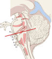
==Pituitary gland== | |||
===Anatomy=== | |||
*Located in sella turcica below optic chiasm. | |||
*Anterior lobe is epithelial. | |||
*Posterior lobe is neuroepithelial. | |||
*The infundibulum connects the pituitary to the brain | |||
Schematic: [[File:Gray1181.png Pituitary gland - Gray's anatomy (WC)]]. | |||
====Images==== | |||
<gallery> | |||
File:Anterior and posterior pituitary.jpg | Anterior & posterior pituitary. (WC) | |||
File:Adenohypofýza HE.jpg | Adenohypophysis. (WC/Držiak) | |||
</gallery> | |||
=See also= | =See also= | ||
Latest revision as of 12:34, 17 October 2022
This article covers basic (normal) neurohistology. It is essential to have a good grasp on neurohistology and neuroanatomy... before doing neuropathology.
This article has some overlap with the neuroanatomy article, as there isn't a clear divider between microscopic and macroscopic.
Normal cells
This section deals with normal cellular constituents of the CNS.
Overview
Central nervous system
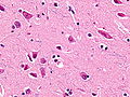
Neuron
- Abundant cytoplasm - key feature.
- Often very large cells, with angled edges.
- Prominent nucleolus.
- Nissl substance (granular perinuclear material = rough endoplasmic reticulum).
Glial cells
- Oligodendrocyte.
- Small round nuclei (lymphocyte-like nucleus) - key feature.
- May resemble a fried egg on H&E (clear cytoplasm, central nucleus).
- Image: oligodendrocyte urmc.rochester.edu
- Astrocyte.
- Irregular non-ovoid nucleus - key feature.
- Nuclei less dense than in oligodendrocyte.
- Close to blood vessels.
- Form blood-brain barrier.
- Cytoplasm normally not visible.
- Image: astrocyte (med.unsw.edu.au) (in endocrine development).
- Microglia - macrophage of the brain (derived from monocyte).
- Typically large cells with abundant cytoplasm.
- Often have vesicles.
- Rarely seen in normal tissue.
- Three morphologic types:
- Ramified microglia.
- Hypertrophic microglia.
- Dystrophic microglia - often seen in neurodegnerative disease.
- Typically large cells with abundant cytoplasm.
- Ependyma.
- Simple ciliated cuboidal epithelium.
- Image: Ependyma (stonybrookmedicalcenter.org).
- Choroid plexus.
- Specialized ependymal cells
- Cuboidal epithelial cells surrounding a core of capillaries and loose connective tissue.
- Ventricular location.
Images

Astrocytes highlighted by GFAP (WC/Jeffery J. Iliff)
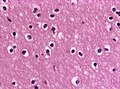
Oligodendrocytes with fried-egg appearance (WC/jensflorian)
Ependymal cells. (WC/marvin101)
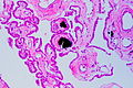
Choroid plexus (WC/Patho)
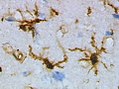
Microglial cells, Lectin stain (WC/Grzegorz Wicher)


Peripheral nervous system
Ganglion cell
- Nerve cell body.
- Found in ganglions - encapsulated body ~ 100-300 μm.
- Large round nucleus with prominent nucleolus - key feature.
- Abundant eosinophilic granular cytoplasm.
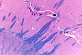
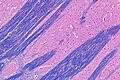
Schwann cell
- Principal glia of the PNS.
- Myelinated or unmyelinated.
- Ovoid, wavy nuclei.
- Cells have a basal membrane.
- Found in peripheral nerves.
- Aka neurolemmocytes.
Images
Ganglion - very high mag. (WC/Nephron)
Peripheral nerve - cross section. (WC/Reytan)
Nerve root HPS stain (WC/Nephron)


WC:
Normal cellular constituents in a table
| Cell | Key feature | Other features | Image |
|---|---|---|---|
| Neuron | cytoplasm | Nissl substance (prominent RER), "sharp" corners in cell membrane, nucleolus - usu. prominent[1] |
red neurons (WC) |
| Astrocyte | non-ovoid nucleus | no cytoplasm | (unsw.edu) |
| Oligodendrocyte | round small nucleus | peri-nuclear clearing | (vetmed.vt.edu) |
| Microglia | rod-like shape, may have "bent" nucleus |
typically have a sharply demarcated bubbly cytoplasm; rarely seen in normal tissue |
(neuropathologyweb.org), (ucsf.edu),(vcu.edu) |
Neurons
There are many types of 'em. Broadly, they can be classified as:
- Pyramidal - have a pyramidal shape.
- Dentrites go to molecular layer.
- Axons go to outside of cortex.
- Non-pyramidal.
Motor neurons:
- Coarse Nissl substance - key feature.
- Nissl described as having a tigroid appearance.[2]
- Polygonal shape.
- Send dendrites in all directions.
Images
www:
Motor neurons - nucleus of CN XII - very high mag. (WC)

Structures
This section deals with structures seen at several places in the CNS.
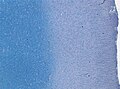
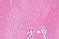
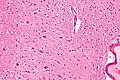
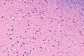
Grey matter and white matter
| Grey matter | White matter | |
| Definition | neurons present | neurons absent |
| Extracellular space (neuropil) |
dense - darker pink (on H&E/HPS) | fine mesh - lighter pink (on H&E/HPS) |
| Image |

Additional images (white matter vs. grey matter):
Grey-white matter interface - high mag. (WC)
Grey and white matter - intermed. mag. (WC)
Normal cortex - LFB. (WC/jensflorian)
White-grey matter junction - LFB. (WC/jensflorian)


Vessels
The small blood vessels in the CNS are separated from the surrounding tissue in histologic sections. This is normal. The spaces are called Virchow-Robin spaces.[3]
Histology by anatomical structure
This section deals with specific anatomical structures.
Subependyma
Features:[4]
- Ependyma (simple ciliated cuboidal epithelium).
- Subependymal plate - connective tissue with blood vessels.
Caudate
Features:
- Neurons with adjacent ependymal lining.[5]
- The caudate forms lateral wall of lateral ventricle.
Notes:
- Caudate, putamen and nucleus accumbens are collectively called neostriatum.[6]
Putamen
Features:
- Striatopallidal fibers AKA pencils of Wilson (also pencil fibers of Wilson[6] and Wilson's pencils[7]) - bundles of blue fibres (on H&E LFB).
- Neurons:
- Small - GABA.
- Large (very rare: ~1 in 100-200) - cholingeric.
Notes:
- Histologically identical to the caudate - but not adjacent to a ventricle.
- The caudate is adjacent to an ependymal lined space, putamen is not.[8]
- Necrotic putamen in methanol poisoning.[9]
Images
Globus pallidus & putamen - showing Wilson's pencils - very low mag. (WC)
Putamen - intermed. mag. (WC)


www:
Globus pallidus
Features:
- Histologically distinct from caudate and putamen.
Image
Globus pallidus, putamen and the nucleus basalis in the substantia innominata - very low mag. (WC)

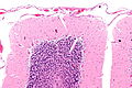
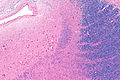
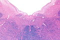
Hippocampus
Structures
Hippocampal formation:[11]
- Dentate gyrus.
- "Dense" thin layer of nuclei.
- Quasi "U-shaped"; "open" (top) portion of "U" is superolateral.
- Image: Dentate gyrus (stonybrookmedicalcenter.org).
- Hippocampus proper (AKA Ammon's horn) - this is subdivided:
- Cornu ammonis 3 (CA3) - location: superior.
- Large pyramidal neurons.
- CA1 (AKA Sommer's sector) - location: inferior (next to subiculum).
- Small dispersed pyramidal neurons.
- CA2 - location: in between CA3 and CA1, lateral.
- Narrow band of neurons between CA3 and CA1.
- CA4 - location: medial (closest to dentate gyrus; CA4 sits in "open" part of "U").
- Cornu ammonis 3 (CA3) - location: superior.
- Subiculum (AKA subicular complex).
- Transitions to the six layers in the entorhinal cortex.
- No vacuolated looking stuff next to it.
- Transitions to the six layers in the entorhinal cortex.
Important notes:
- CA1 - weak link, dies in ischemia, affected by hypoglycemia, degenerative diseases and toxins.
- CA2 - resistant to ischemia.
- CA4 - involved in epilepsy,[12] usu. normal in degenerative diseases.[13]
Images
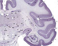
Hippocampus - frontal section. (WC)
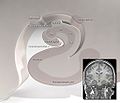
Hippocampus - schematic. (WC)

.jpg)
www:
- Hippocampus (ajnr.org).
- Hippocampus and subiculum (hu-berlin.de).
- Hippocampus - crappy schematic (ucsd.edu).
Layers of CA[14]
- Molecular layer - opposed to the dentate gyrus (of Hippocampal formation).
- Neurons (described above).
- Alveus - opposed to the lateral ventricle.
- Connects to the mammillary bodies via the fornix (circuit of Papez).
Cerebellum
Main components:
- Cortex (superficial) - branches (Christmas tree-like).
- Dentate nucleus (deep) - looks like the bite impression of a molar.
Dentate nucleus
Features:[15]
- Ribbon of grey matter.
- Large neurons.
- Small neurons.
Images
Dentate nucleus of the cerebellum - intermed. mag. (WC)
Dentate nucleus of the cerebellum - high mag. (WC)


Cerebellar cortex
- Layers (superficial to deep) - mnemonic MPG:[16]
- Molecular layer -- "very pink" on H&E.
- Inhibitory interneurons: stellate cells, basket cells.
- Purkinje cell layer.
- One cell layer thick - hueuege cells (~50-80 micrometers[1]).
- Very large nucleus (~4x RBC diameter =~ 4x the size of granule cell).
- Large nucleolus (~1x RBC diameter =~ size of granule cell).
- Very large nucleus (~4x RBC diameter =~ 4x the size of granule cell).
- One cell layer thick - hueuege cells (~50-80 micrometers[1]).
- Granule cell layer -- "very blue" on H&E.
- Granule cells (neurons) - majority of cells, small (~10 micrometres), round.
- May look like small cell carcinoma to the uninitiated.
- Golgi cells (interneurons) - few in number, elongated/columnar, 3-5x size of granule cell.
- Granule cells (neurons) - majority of cells, small (~10 micrometres), round.
- Molecular layer -- "very pink" on H&E.
Notes:
- Bergmann glia are found between the molecular layer & granular layer. They are normally not seen. They are increased & prominent in pathologic states (e.g. ischemia); "Bergmann gliosis".[17]
Images
www:
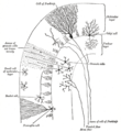
Cerebellar cortex - schematic. (Gray's Anatomy/WC)
Cerebellar cortex - intermed. mag. (WC)
Cerebellar cortex - high mag. Bielschowsky stain. (WC)


Cerebral cortex
Layers (superficial to deep):
- Molecular layer.
- Empty appearing.
- Outer granular layer.
- Higher cell density & smaller cells than pyramidal layer.
- Outer pyramidal layer.
- Inner granular layer.
- Not prominent in frontal cortex.
- Where the thalamic axons end.
- Divided in three (a, b, c) in the calcarine cortex due to two white matter bands (external band of Baillarger, internal band of Baillarger) that are grossly identified as the line of Gennari.[18][19]
- Image: Calcarine cortex (ouhsc.edu).[20]
- Inner pyramidal layer.
- Location of Betz neurons - large motor neurons of cerebral cortex.
- Multiforme layer (Polymorphic layer).
Images:
- Cajal drawings - different areas (WC).
- Visual cortex - intermed. mag. (WC).
- Visual cortex - nissl stain (nlm.nih.gov).[21]
- Different stains (rice.edu).
- Cerebral cortex (williamcalvin.com).
- Cerebral cortex (benbest.com).
Cingulate cortex
- Spindle neurons, AKA von Economo neurons.
- Thought to be important in cognition and problem solving.[22]
Images
Spindle neurons - high mag. (WC)
Spindle neurons - cropped - very high mag. (WC)


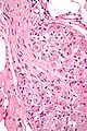
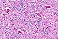
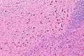
Pineal gland
- Cells in lobulated clusters or linear arrays (low power).
- Pinealocyte:
- Light staining and round nuclei with neuroendocrine look (i.e. salt-and-pepper chromatin).
- Broad rim of light cytoplasm.
- Astrocytes:
- Cylindrical hyperchromatic nucleus ~ 1/2 the size of pinealocyte.
Notes:
- Highly cellular structure - may be confused with (metastatic) small cell carcinoma.
- Often calcified.
Images
Pineal gland - very high mag. (WC/Nephron)
Pineal gland - intermed. mag. (WC/Nephron)
Pineal gland. (WC)


www:
IHC
- Synaptophysin +ve.[23]
Midbrain
Structures:
- Substantia nigra (Parkinson's disease).
- Nuclei of CN IV (posterior).
- Nuclei of CN III (anterior).
- Cerebral penduncles (anterior).
- Red nuclei.
Schematics
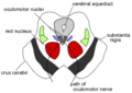
Midbrain (WC)
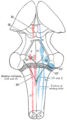
Nuclei of the CNs (WC)
Nuclei of the CNs - sagittal section of brain stem (WC)


Pons
Features:
- Looks like bacon (at very low power).[25]
- Images:
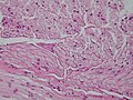
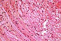
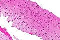
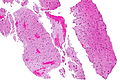
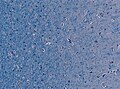
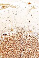
Locus ceruleus
- Literally means blue spot.
- Location: adjacent to midline + anterior to 4th ventricle.
Microscopic features:
- Pigmented neurons.
- Produce norepinephrine.
Notes:
- Pale in Parkinson disease due to neuronal loss.[26]
Images
Locus ceruleus (LC) - very low mag. (WC)
Locus ceruleus (LC) - low mag. (WC)
Locus ceruleus (LC) - intermed. mag. (WC)
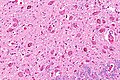
Locus ceruleus (LC) - high mag. (WC)


www:
Medulla oblongata
- AKA medulla.
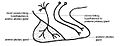
Anatomy
Schematic: Medulla oblongata - Gray's anatomy (WC).
Anterior
- Pyramids: adjacent to midline, anterior.
- Olives: lateral and posterior to pyramids.
Posterior - important nuclei (location)
- CN XII: 4th ventricle + adjacent to midline; medial to nucleus of CN X.[27]
- CN X: 4th ventricle + lateral to nucleus of CN XII.
Image
Medulla oblongata - very low mag. (WC)

Pituitary gland
Anatomy
- Located in sella turcica below optic chiasm.
- Anterior lobe is epithelial.
- Posterior lobe is neuroepithelial.
- The infundibulum connects the pituitary to the brain
Schematic: File:Gray1181.png Pituitary gland - Gray's anatomy (WC).
Images
Anterior & posterior pituitary. (WC)
Adenohypophysis. (WC/Držiak)

See also
References
- ↑ 1.0 1.1 Perry, Arie; Brat, Daniel J. (2010). Practical Surgical Neuropathology: A Diagnostic Approach: A Volume in the Pattern Recognition series (1st ed.). Churchill Livingstone. pp. 16. ISBN 978-0443069826.
- ↑ URL: http://www.stonybrookmedicalcenter.org/pathology/neuropathology/chapter1. Accessed on: 5 July 2010.
- ↑ URL: http://www.whonamedit.com/synd.cfm/43.html. Accessed on: 2 August 2011.
- ↑ Croul SE. 28 June 2010.
- ↑ URL: http://www.stonybrookmedicalcenter.org/pathology/neuropathology/chapter1. Accessed on: 2 July 2010.
- ↑ 6.0 6.1 Perry, Arie; Brat, Daniel J. (2010). Practical Surgical Neuropathology: A Diagnostic Approach: A Volume in the Pattern Recognition series (1st ed.). Churchill Livingstone. pp. 23-34. ISBN 978-0443069826.
- ↑ Kimura M, Kato M, Shimazaki H, Watanabe K, Matsumoto N (December 1996). "Neural information transferred from the putamen to the globus pallidus during learned movement in the monkey". J. Neurophysiol. 76 (6): 3771–86. PMID 8985875. http://jn.physiology.org/cgi/pmidlookup?view=long&pmid=8985875.
- ↑ URL: http://www.stonybrookmedicalcenter.org/pathology/neuropathology/chapter1. Accessed on: 22 December 2010.
- ↑ Bhatia, R.; Kumar, M.; Garg, A.; Nanda, A. (Dec 2008). "Putaminal necrosis due to methanol toxicity.". Pract Neurol 8 (6): 386-7. doi:10.1136/jnnp.2008.161976. PMID 19015300.
- ↑ Perry, Arie; Brat, Daniel J. (2010). Practical Surgical Neuropathology: A Diagnostic Approach: A Volume in the Pattern Recognition series (1st ed.). Churchill Livingstone. pp. 23. ISBN 978-0443069826.
- ↑ URL: http://www.stonybrookmedicalcenter.org/pathology/neuropathology/chapter1. Accessed on: 2 July 2010.
- ↑ Ingvar, M. (1986). "Cerebral blood flow and metabolic rate during seizures. Relationship to epileptic brain damage.". Ann N Y Acad Sci 462: 194-206. PMID 3518570.
- ↑ D. Munoz. 27 July 2011.
- ↑ Perry, Arie; Brat, Daniel J. (2010). Practical Surgical Neuropathology: A Diagnostic Approach: A Volume in the Pattern Recognition series (1st ed.). Churchill Livingstone. pp. 25. ISBN 978-0443069826.
- ↑ Perry, Arie; Brat, Daniel J. (2010). Practical Surgical Neuropathology: A Diagnostic Approach: A Volume in the Pattern Recognition series (1st ed.). Churchill Livingstone. pp. 27. ISBN 978-0443069826.
- ↑ URL: http://www.stonybrookmedicalcenter.org/pathology/neuropathology/chapter1. Accessed on: 2 July 2010.
- ↑ Perry, Arie; Brat, Daniel J. (2010). Practical Surgical Neuropathology: A Diagnostic Approach: A Volume in the Pattern Recognition series (1st ed.). Churchill Livingstone. pp. 18. ISBN 978-0443069826.
- ↑ Perry, Arie; Brat, Daniel J. (2010). Practical Surgical Neuropathology: A Diagnostic Approach: A Volume in the Pattern Recognition series (1st ed.). Churchill Livingstone. pp. 24. ISBN 978-0443069826.
- ↑ URL: http://www.ncbi.nlm.nih.gov/books/NBK11524/. Accessed on: 7 January 2011.
- ↑ URL: http://moon.ouhsc.edu/kfung/iacp-olp/apaq-text/N1-MS-01-01-Ans.htm and http://moon.ouhsc.edu/kfung/iacp-olp/apaq-text/n1-ms-01.htm. Accessed on: 31 October 2010.
- ↑ URL: http://www.ncbi.nlm.nih.gov/books/NBK11524/. Accessed on: 7 January 2011.
- ↑ Allman, JM.; Hakeem, A.; Erwin, JM.; Nimchinsky, E.; Hof, P. (May 2001). "The anterior cingulate cortex. The evolution of an interface between emotion and cognition.". Ann N Y Acad Sci 935: 107-17. PMID 11411161.
- ↑ 23.0 23.1 Perry, Arie; Brat, Daniel J. (2010). Practical Surgical Neuropathology: A Diagnostic Approach: A Volume in the Pattern Recognition series (1st ed.). Churchill Livingstone. pp. 25-26. ISBN 978-0443069826.
- ↑ 24.0 24.1 URL: http://www.lab.anhb.uwa.edu.au/mb140/corepages/endocrines/endocrin.htm. Accessed on: 31 October 2010.
- ↑ Croul SE. 28 June 2010.
- ↑ Mitchell, Richard; Kumar, Vinay; Fausto, Nelson; Abbas, Abul K.; Aster, Jon (2011). Pocket Companion to Robbins & Cotran Pathologic Basis of Disease (8th ed.). Elsevier Saunders. pp. 677. ISBN 978-1416054542.
- ↑ Crossman, Alan R.; Neary, David. (2010). Neuroanatomy: An Illustrated Colour Text (4th ed.). Churchill Livingstone. pp. 13. ISBN 978-0702030864.
- ↑ Miller VM, Kenny RA, Oakley AE, Hall R, Kalaria RN, Allan LM (August 2009). "Dorsal motor nucleus of vagus protein aggregates in Lewy body disease with autonomic dysfunction". Brain Res. 1286: 165–73. doi:10.1016/j.brainres.2009.05.083. PMID 19501577.