Nasopharyngeal angiofibroma
Jump to navigation
Jump to search
The printable version is no longer supported and may have rendering errors. Please update your browser bookmarks and please use the default browser print function instead.
| Nasopharyngeal angiofibroma | |
|---|---|
| Diagnosis in short | |
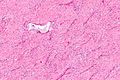
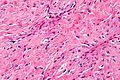
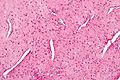
 Nasopharyngeal angiofibroma. H&E stain. | |
|
| |
| LM | fibroblastic cells with plump (near cuboidal) nuclei, fibrous stroma, abundant capillaries |
| Site | head and neck - nasopharynx |
|
| |
| Clinical history | male, adolescents young to adults, frequent nose bleeds |
| Prevalence | uncommon |
| Prognosis | benign |
| Treatment | surgical resection |
Nasopharyngeal angiofibroma is a benign lesion of the head and neck.
General
- Classically adolescent males with recurrent nose bleeds.
- Age range 8-41 in one series of 162 cases.[1]
- Can be subtyped by location into three groups.[2]
Microscopic
Features:[3]
- Fibroblastic cells with plump (near cuboidal) nuclei.
- Fibrous stroma.
- Abundant capillaries.
Images
Intermed. mag.

High mag.
Very high mag.


High mag.
See also
References
- ↑ Huang, Y.; Liu, Z.; Wang, J.; Sun, X.; Yang, L.; Wang, D. (Aug 2014). "Surgical management of juvenile nasopharyngeal angiofibroma: analysis of 162 cases from 1995 to 2012.". Laryngoscope 124 (8): 1942-6. doi:10.1002/lary.24522. PMID 24222212.
- ↑ Yi, Z.; Fang, Z.; Lin, G.; Lin, C.; Xiao, W.; Li, Z.; Cheng, J.; Zhou, A.. "Nasopharyngeal angiofibroma: a concise classification system and appropriate treatment options.". Am J Otolaryngol 34 (2): 133-41. doi:10.1016/j.amjoto.2012.10.004. PMID 23332298.
- ↑ Klatt, Edward C. (2006). Robbins and Cotran Atlas of Pathology (1st ed.). Saunders. pp. 144. ISBN 978-1416002741.