Difference between revisions of "Mucinous cystadenoma of the ovary"
Jump to navigation
Jump to search
| (16 intermediate revisions by the same user not shown) | |||
| Line 11: | Line 11: | ||
| IHC = | | IHC = | ||
| EM = | | EM = | ||
| Molecular = | | Molecular = +/-[[KRAS mutation]] | ||
| IF = | | IF = | ||
| Gross = multiloculated cystic lesion, thin cyst walls/no solid areas | | Gross = multiloculated cystic lesion, thin cyst walls/no solid areas | ||
| Line 27: | Line 27: | ||
| Prognosis = benign | | Prognosis = benign | ||
| Other = | | Other = | ||
| ClinDDx = | | ClinDDx = other [[ovarian tumours]] - esp. mucinous ones, and mucinous appendiceal lesions | ||
| Tx = cystectomy/oophorectomy | | Tx = cystectomy/oophorectomy | ||
}} | }} | ||
| Line 36: | Line 36: | ||
==General== | ==General== | ||
*Common.<ref name=pmid22246404>{{Cite journal | last1 = Pongsuvareeyakul | first1 = T. | last2 = Khunamornpong | first2 = S. | last3 = Settakorn | first3 = J. | last4 = Sukpan | first4 = K. | last5 = Suprasert | first5 = P. | last6 = Siriaunkgul | first6 = S. | title = Accuracy of frozen-section diagnosis of ovarian mucinous tumors. | journal = Int J Gynecol Cancer | volume = 22 | issue = 3 | pages = 400-6 | month = Mar | year = 2012 | doi = 10.1097/IGC.0b013e31823dc328 | PMID = 22246404 }}</ref> | *Common.<ref name=pmid22246404>{{Cite journal | last1 = Pongsuvareeyakul | first1 = T. | last2 = Khunamornpong | first2 = S. | last3 = Settakorn | first3 = J. | last4 = Sukpan | first4 = K. | last5 = Suprasert | first5 = P. | last6 = Siriaunkgul | first6 = S. | title = Accuracy of frozen-section diagnosis of ovarian mucinous tumors. | journal = Int J Gynecol Cancer | volume = 22 | issue = 3 | pages = 400-6 | month = Mar | year = 2012 | doi = 10.1097/IGC.0b013e31823dc328 | PMID = 22246404 }}</ref> | ||
**Approximately 80% of all mucinous ovarian tumours.<ref name=pmid15626914>{{cite journal |author=Hart WR |title=Mucinous tumors of the ovary: a review |journal=Int. J. Gynecol. Pathol. |volume=24 |issue=1 |pages=4–25 |year=2005 |month=January |pmid=15626914 |doi= |url=}}</ref> | |||
*Benign. | *Benign. | ||
| Line 42: | Line 43: | ||
*May be very large large.<ref name=pmid22283565>{{Cite journal | last1 = Leys | first1 = CM. | last2 = Gasior | first2 = AC. | last3 = Hornberger | first3 = LL. | last4 = St Peter | first4 = SD. | title = Laparoscopic resection of massive ovarian mucinous cystadenoma. | journal = J Laparoendosc Adv Surg Tech A | volume = 22 | issue = 3 | pages = 307-10 | month = Apr | year = 2012 | doi = 10.1089/lap.2011.0435 | PMID = 22283565 }}</ref> | *May be very large large.<ref name=pmid22283565>{{Cite journal | last1 = Leys | first1 = CM. | last2 = Gasior | first2 = AC. | last3 = Hornberger | first3 = LL. | last4 = St Peter | first4 = SD. | title = Laparoscopic resection of massive ovarian mucinous cystadenoma. | journal = J Laparoendosc Adv Surg Tech A | volume = 22 | issue = 3 | pages = 307-10 | month = Apr | year = 2012 | doi = 10.1089/lap.2011.0435 | PMID = 22283565 }}</ref> | ||
*No solid areas/thin cyst walls. | *No solid areas/thin cyst walls. | ||
===Images=== | |||
<gallery> | |||
Image:Mucinous Cystadenoma of Ovary (218889489).jpg | Mucinous cystadenoma of the ovary. (WC) | |||
</gallery> | |||
==Microscopic== | ==Microscopic== | ||
| Line 47: | Line 53: | ||
*Cysts lined by a simple mucinous epithelium. | *Cysts lined by a simple mucinous epithelium. | ||
*No cytologic atypia. | *No cytologic atypia. | ||
Note: | |||
*A borderline component may be present but ''must be <10%'' of the tumour.<ref name=Ref_GP>{{Ref GP|416}}</ref> | |||
**Lesions with <10% borderline component are known as ''mucinous cystadenoma of the ovary with focal proliferation'' or ''mucinous cystadenoma of the ovary with focal atypia''. | |||
DDx: | DDx: | ||
| Line 65: | Line 75: | ||
Image: Ovarian mucinous cystadenoma - a3- very high mag.jpg | OMC - very high mag. | Image: Ovarian mucinous cystadenoma - a3- very high mag.jpg | OMC - very high mag. | ||
</gallery> | </gallery> | ||
==Molecular== | |||
*[[KRAS mutation]]s common ~ 56% of mucinous cystadenomas in one series.<ref name=pmid9118042>{{Cite journal | last1 = Cuatrecasas | first1 = M. | last2 = Villanueva | first2 = A. | last3 = Matias-Guiu | first3 = X. | last4 = Prat | first4 = J. | title = K-ras mutations in mucinous ovarian tumors: a clinicopathologic and molecular study of 95 cases. | journal = Cancer | volume = 79 | issue = 8 | pages = 1581-6 | month = Apr | year = 1997 | doi = | PMID = 9118042 }}</ref> | |||
==Sign out== | |||
<pre> | |||
Left Ovarian Cyst, Excision: | |||
- Mucinous cystadenoma. | |||
</pre> | |||
===Mucinous cystadenoma with focal proliferation=== | |||
<pre> | |||
Left Ovarian Cyst, Excision: | |||
- Mucinous cystadenoma with focal proliferation, see comment. | |||
Comment: | |||
The proliferative component is less than 10% of the lesion. | |||
</pre> | |||
==See also== | ==See also== | ||
*[[Ovarian tumours]]. | *[[Ovarian tumours]]. | ||
*[[Mucinous ovarian tumours]]. | |||
*[[Mucinous cystadenoma]]. | *[[Mucinous cystadenoma]]. | ||
*[[Mucinous cystadenoma of the appendix]]. | *[[Mucinous cystadenoma of the appendix]]. | ||
Latest revision as of 16:36, 1 March 2018
| Mucinous cystadenoma of the ovary | |
|---|---|
| Diagnosis in short | |
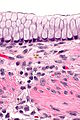
 Ovarian mucinous cystadenoma. H&E stain. | |
|
| |
| LM | cysts lined by a simple mucinous epithelium, no cytologic atypia |
| LM DDx | seromucinous borderline tumour of the ovary, mucinous borderline tumour of the ovary |
| Molecular | +/-KRAS mutation |
| Gross | multiloculated cystic lesion, thin cyst walls/no solid areas |
| Site | ovary - see ovarian tumours |
|
| |
| Prevalence | common |
| Prognosis | benign |
| Clin. DDx | other ovarian tumours - esp. mucinous ones, and mucinous appendiceal lesions |
| Treatment | cystectomy/oophorectomy |
Mucinous cystadenoma of the ovary, also ovarian mucinous cystadenoma, is a common benign ovarian tumour.
It is unrelated to mucinous cystadenoma of the appendix.
General
Gross
- Usually multiloculated.
- May be very large large.[3]
- No solid areas/thin cyst walls.
Images
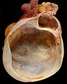
Mucinous cystadenoma of the ovary. (WC)
.jpg)
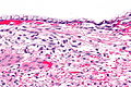
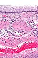
Microscopic
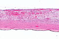
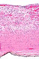
Features:
- Cysts lined by a simple mucinous epithelium.
- No cytologic atypia.
Note:
- A borderline component may be present but must be <10% of the tumour.[4]
- Lesions with <10% borderline component are known as mucinous cystadenoma of the ovary with focal proliferation or mucinous cystadenoma of the ovary with focal atypia.
DDx:
Images
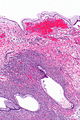
OMC - low mag.
OMC - low mag.
OMC - intermed. mag.
OMC - high mag.
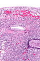
OMC - intermed. mag.
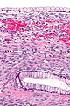
OMC - high mag.
OMC - high mag.
OMC - very high mag.


Molecular
- KRAS mutations common ~ 56% of mucinous cystadenomas in one series.[5]
Sign out
Left Ovarian Cyst, Excision:
	- Mucinous cystadenoma.
Mucinous cystadenoma with focal proliferation
Left Ovarian Cyst, Excision:
	- Mucinous cystadenoma with focal proliferation, see comment.

Comment:
The proliferative component is less than 10% of the lesion.
See also
- Ovarian tumours.
- Mucinous ovarian tumours.
- Mucinous cystadenoma.
- Mucinous cystadenoma of the appendix.
References
- ↑ Pongsuvareeyakul, T.; Khunamornpong, S.; Settakorn, J.; Sukpan, K.; Suprasert, P.; Siriaunkgul, S. (Mar 2012). "Accuracy of frozen-section diagnosis of ovarian mucinous tumors.". Int J Gynecol Cancer 22 (3): 400-6. doi:10.1097/IGC.0b013e31823dc328. PMID 22246404.
- ↑ Hart WR (January 2005). "Mucinous tumors of the ovary: a review". Int. J. Gynecol. Pathol. 24 (1): 4–25. PMID 15626914.
- ↑ Leys, CM.; Gasior, AC.; Hornberger, LL.; St Peter, SD. (Apr 2012). "Laparoscopic resection of massive ovarian mucinous cystadenoma.". J Laparoendosc Adv Surg Tech A 22 (3): 307-10. doi:10.1089/lap.2011.0435. PMID 22283565.
- ↑ Nucci, Marisa R.; Oliva, Esther (2009). Gynecologic Pathology: A Volume in Foundations in Diagnostic Pathology Series (1st ed.). Churchill Livingstone. pp. 416. ISBN 978-0443069208.
- ↑ Cuatrecasas, M.; Villanueva, A.; Matias-Guiu, X.; Prat, J. (Apr 1997). "K-ras mutations in mucinous ovarian tumors: a clinicopathologic and molecular study of 95 cases.". Cancer 79 (8): 1581-6. PMID 9118042.
.