Difference between revisions of "Mesonephric duct remnant"
Jump to navigation
Jump to search
(=images) |
(→Microscopic: +images) |
||
| Line 36: | Line 36: | ||
*[[Wolffian duct hyperplasia]]. | *[[Wolffian duct hyperplasia]]. | ||
Images: | ==Images== | ||
<gallery> | |||
Image: Mesonephric duct remnant -- very low mag.jpg | MDR - very low mag. | |||
Image: Mesonephric duct remnant -- low mag.jpg | MDR - low mag. | |||
Image: Mesonephric duct remnant -- intermed mag.jpg | MDR - intermed. mag. | |||
Image: Mesonephric duct remnant -- high mag.jpg | MDR - high mag. | |||
Image: Mesonephric duct remnant -- very high mag.jpg | MDR - very high mag. | |||
Image: Mesonephric duct remnant - alt -- very high mag.jpg | MDR - very high mag. | |||
</gallery> | |||
www: | |||
*[http://www.webpathology.com/image.asp?case=550&n=1 Mesonephric duct remnant - low mag. (webpathology.com)]. | *[http://www.webpathology.com/image.asp?case=550&n=1 Mesonephric duct remnant - low mag. (webpathology.com)]. | ||
*[http://www.webpathology.com/image.asp?n=2&Case=550 Mesonephric duct remnant - high mag. (webpathology.com)]. | *[http://www.webpathology.com/image.asp?n=2&Case=550 Mesonephric duct remnant - high mag. (webpathology.com)]. | ||
Revision as of 05:21, 14 December 2015

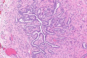
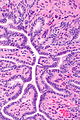
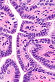
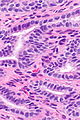
Micrograph showing the mesonephric duct remnant. H&E stain.
Mesonephric duct remnant, also Wolffian duct remnant and Gartner duct, a benign embryological remanant that is usually ignored.
Gartner duct cyst, mesonephric duct cyst and Wolffian duct cyst redirect here.
General
Epidemiology:
- Embryological remnant - benign.
- Wolffian duct = precursor of male reproductive tract.[1]
Notes:
- This is not a finding that is reported. The importance of this finding is knowing it isn't something neoplastic.
- Lame way of remember the synonyms Gartner, Mesonephric, and Wolffian: GMW... it is trying to be a BMW but in girls.
Gross
Note:
- Same location as Muellerian cyst.
Images

Schematic showing the location and development of the MDR. (WC/Gray's Anatomy)
Microscopic
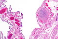
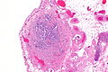
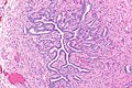
Features:[3]
- Small duct -- typically ~50-100 micrometres in diameter.
- Duct lined by cuboidal cells with moderate eosinophilic cytoplasm.
DDx:
- Muellerian cyst - most common vaginal cyst.[2]
- Vaginal inclusion cyst.
- Cervical adenocarcinoma, not otherwise specified.
- Minimal deviation adenocarcinoma of the uterine cervix.
- Mesonephric adenocarcinoma - has cellular atypia.
- Wolffian duct hyperplasia.
Images
MDR - very low mag.
MDR - low mag.
MDR - intermed. mag.
MDR - high mag.
MDR - very high mag.
MDR - very high mag.


www:
- Mesonephric duct remnant - low mag. (webpathology.com).
- Mesonephric duct remnant - high mag. (webpathology.com).
IHC
Features:[4]
- CD10 +ve.
- CK7 +ve.
- PAX2 +ve (nuclear - strong & diffuse).[5]
- Cancers often lose staining.
See also
References
- ↑ Hannema SE, Print CG, Charnock-Jones DS, Coleman N, Hughes IA (2006). "Changes in gene expression during Wolffian duct development". Horm. Res. 65 (4): 200–9. doi:10.1159/000092408. PMID 16567946.
- ↑ 2.0 2.1 Nucci, Marisa R.; Oliva, Esther (2009). Gynecologic Pathology: A Volume in Foundations in Diagnostic Pathology Series (1st ed.). Churchill Livingstone. pp. 97. ISBN 978-0443069208.
- ↑ Sternberg SE. Histology for Pathologists. 2nd Ed. P.893.
- ↑ Humphrey, Peter A; Dehner, Louis P; Pfeifer, John D (2008). The Washington Manual of Surgical Pathology (1st ed.). Lippincott Williams & Wilkins. pp. 442. ISBN 978-0781765275.
- ↑ Rabban, JT.; McAlhany, S.; Lerwill, MF.; Grenert, JP.; Zaloudek, CJ. (Feb 2010). "PAX2 distinguishes benign mesonephric and mullerian glandular lesions of the cervix from endocervical adenocarcinoma, including minimal deviation adenocarcinoma.". Am J Surg Pathol 34 (2): 137-46. doi:10.1097/PAS.0b013e3181c89c98. PMID 20061933.