Difference between revisions of "Merkel cell carcinoma"
Jump to navigation
Jump to search
(redirect) |
m (many ALK1 +ve) |
||
| (14 intermediate revisions by one other user not shown) | |||
| Line 1: | Line 1: | ||
{{ Infobox diagnosis | |||
| Name = {{PAGENAME}} | |||
| Image = Merkel_cell_carcinoma_-_high_mag.jpg | |||
| Width = | |||
| Caption = Merkel cell carcinoma. [[H&E stain]]. | |||
| Micro = neuroendocrine nuclear features (round nucleus, small nucleoli/no nucleolus, stippled chromatin), usually scant cytoplasm, usually small (~3x resting lymphocyte), often in sheets | |||
| Subtypes = | |||
| LMDDx = [[small cell carcinoma]], cutaneous [[Ewing sarcoma]], [[Burkitt lymphoma]], other [[small round blue cell tumours]] | |||
| Stains = | |||
| IHC = Merkel cell polyomavirus +ve, CK20 +ve (perinuclear dot-like), CD56 +ve, TTF-1 -ve, CK7 -ve, NF +ve | |||
| EM = | |||
| Molecular = | |||
| IF = | |||
| Gross = | |||
| Grossing = | |||
| Site = [[skin]] | |||
| Assdx = | |||
| Syndromes = | |||
| Clinicalhx = +/-immunosuppressed/immunoincompetent | |||
| Signs = | |||
| Symptoms = | |||
| Prevalence = rare | |||
| Bloodwork = | |||
| Rads = | |||
| Endoscopy = | |||
| Prognosis = poor | |||
| Other = | |||
| ClinDDx = | |||
}} | |||
{{ Infobox external links | |||
| Name = {{PAGENAME}} | |||
| EHVSC = | |||
| EHVSC_mult = | |||
| pathprotocols = | |||
| wikipedia = merkel cell carcinoma | |||
| pathoutlines = | |||
}} | |||
'''Merkel cell carcinoma''', abbreviated '''MCC''', is an uncommon aggressive form of skin cancer. | |||
==General== | |||
Features:<ref name=pmid20418670>{{Cite journal | last1 = Calder | first1 = KB. | last2 = Smoller | first2 = BR. | title = New insights into merkel cell carcinoma. | journal = Adv Anat Pathol | volume = 17 | issue = 3 | pages = 155-61 | month = May | year = 2010 | doi = 10.1097/PAP.0b013e3181d97836 | PMID = 20418670 }}</ref> | |||
*Rare. | |||
*Aggressive course/poor prognosis. | |||
*Neuroendocrine-like.<ref name=pmid19395876>{{Cite journal | last1 = Pulitzer | first1 = MP. | last2 = Amin | first2 = BD. | last3 = Busam | first3 = KJ. | title = Merkel cell carcinoma: review. | journal = Adv Anat Pathol | volume = 16 | issue = 3 | pages = 135-44 | month = May | year = 2009 | doi = 10.1097/PAP.0b013e3181a12f5a | PMID = 19395876 }} | |||
</ref> | |||
Etiology: | |||
*Most caused by ''Merkel cell [[polyomavirus]]''.<ref name=pmid18202256>{{Cite journal | last1 = Feng | first1 = H. | last2 = Shuda | first2 = M. | last3 = Chang | first3 = Y. | last4 = Moore | first4 = PS. | title = Clonal integration of a polyomavirus in human Merkel cell carcinoma. | journal = Science | volume = 319 | issue = 5866 | pages = 1096-100 | month = Feb | year = 2008 | doi = 10.1126/science.1152586 | PMID = 18202256 }}</ref><ref name=pmid20418670/> | |||
*Immunocompromised/immunosuppressed (e.g. organ transplant recipients). | |||
==Microscopic== | |||
Features:<ref name=Ref_WMSP491>{{Ref WMSP|491}}</ref> | |||
*Neuroendocrine nuclear features - round nucleus, small nucleoli/no nucleolus, stippled chromatin - '''key feature'''. | |||
*Typically medium size cells ~3x resting lymphocyte. | |||
**May be small or large. | |||
*Architecture: nests, sheets or trabeculae. | |||
*Scant cytoplasm. | |||
*Abundant mitoses. † | |||
*+/-Nuclear moulding. | |||
**Nuclei of adjacent cells conform to one another. | |||
*+/-Tumour infiltrating lymphocytes. ‡ | |||
Notes: | |||
*† >10 mitoses/HPF = poor prognosis - definition suffers from [[HPFitis]].<ref name=capp_mcc>URL: [http://www.cap.org/apps/docs/committees/cancer/cancer_protocols /2011/SkinMerkelCell_11protocol.pdf http://www.cap.org/apps/docs/committees/cancer/cancer_protocols/2011/SkinMerkelCell_11protocol.pdf]. Accessed on: 28 March 2012.</ref> | |||
*‡ May be associated with a worse prognosis.<ref name=capp_mcc/> | |||
*Merkel cell carcinoma [[lymph node metastases]] is difficult to diagnose with routine stains; use of IHC stains are advised.<ref name=capp_mcc/> | |||
*Arise from the epidermis - very rarely in situ.<ref name=pmid15606676>{{Cite journal | last1 = Ferringer | first1 = T. | last2 = Rogers | first2 = HC. | last3 = Metcalf | first3 = JS. | title = Merkel cell carcinoma in situ. | journal = J Cutan Pathol | volume = 32 | issue = 2 | pages = 162-5 | month = Feb | year = 2005 | doi = 10.1111/j.0303-6987.2005.00270.x | PMID = 15606676 }}</ref> | |||
DDx: | |||
*[[Basal cell carcinoma]] - no stippled chromatin, less mitoses active. | |||
*Cutaneous [[Ewing sarcoma]] - sorted-out with immunostains. | |||
*[[Lymphoma]]. | |||
**[[Burkitt lymphoma]]. | |||
*Metastatic [[small cell carcinoma]]. | |||
*Other [[small round cell tumours]]. | |||
===Images=== | |||
<gallery> | |||
Image:Merkel_cell_carcinoma_-_intermed_mag.jpg | MCC - intermed. mag. (WC/Nephron) | |||
Image:Merkel_cell_carcinoma_-_high_mag.jpg | MCC - high mag. (WC/Nephron) | |||
Image:Merkel_cell_carcinoma_-_very_high_mag.jpg | MCC - very high mag. (WC/Nephron) | |||
Image:Merkelcellcarcinoma_Tag.jpg | Merkel cell carcinoma - nested pattern (WC) | |||
</gallery> | |||
www: | |||
*[http://www.bccancer.bc.ca/HPI/CE/cytotechnology/cytosleuthquiz/nongyne/ng12hist.htm MCC (bccancer.bc.ca)]. | |||
*[http://www.joplink.net/prev/200403/07.html MCC (joplink.net)]. | |||
*[http://www.ispub.com/ispub/ijd/volume_5_number_2_8/concurrent_merkel_cell_carcinoma_and_bowen_s_disease_of_the_thigh/bowen-fig3.jpg Merkel cell carcinoma (ispub.com)]. | |||
*[http://path.upmc.edu/cases/case398.html Merkel cell carcinoma - several images (upmc.edu)]. | |||
==IHC== | |||
Features: | |||
*CK7 -ve. | |||
*CK20 +ve (perinuclear dot-like).<ref name=pmid11533085>{{Cite journal | last1 = Leech | first1 = SN. | last2 = Kolar | first2 = AJ. | last3 = Barrett | first3 = PD. | last4 = Sinclair | first4 = SA. | last5 = Leonard | first5 = N. | title = Merkel cell carcinoma can be distinguished from metastatic small cell carcinoma using antibodies to cytokeratin 20 and thyroid transcription factor 1. | journal = J Clin Pathol | volume = 54 | issue = 9 | pages = 727-9 | month = Sep | year = 2001 | doi = | PMID = 11533085 | URL = http://www.ncbi.nlm.nih.gov/pmc/articles/PMC1731517/ }}</ref> | |||
*CAM5.2 +ve (dot-like pattern). | |||
*CD56 +ve. | |||
*AE1/AE3 +ve. | |||
*Merkel cell polyomavirus +ve ~85% of cases.<ref name=pmid21870327>{{Cite journal | last1 = Jung | first1 = HS. | last2 = Choi | first2 = YL. | last3 = Choi | first3 = JS. | last4 = Roh | first4 = JH. | last5 = Pyon | first5 = JK. | last6 = Woo | first6 = KJ. | last7 = Lee | first7 = EH. | last8 = Jang | first8 = KT. | last9 = Han | first9 = J. | title = Detection of Merkel cell polyomavirus in Merkel cell carcinomas and small cell carcinomas by PCR and immunohistochemistry. | journal = Histol Histopathol | volume = 26 | issue = 10 | pages = 1231-41 | month = Oct | year = 2011 | doi = | PMID = 21870327 }}</ref> | |||
*NF +ve (12 of 13 cases).<ref name=pmid16625069>{{cite journal |authors=Bobos M, Hytiroglou P, Kostopoulos I, Karkavelas G, Papadimitriou CS |title=Immunohistochemical distinction between merkel cell carcinoma and small cell carcinoma of the lung |journal=Am J Dermatopathol |volume=28 |issue=2 |pages=99–104 |date=April 2006 |pmid=16625069 |doi=10.1097/01.dad.0000183701.67366.c7 |url=}}</ref> | |||
**Useful to differentiate from [[small cell carcinoma of lung]]. | |||
Others: | |||
*TTF-1 -ve. | |||
*NSE +ve.<ref name=pmid15606676/> | |||
*PAX5 +ve.<ref name=pmid25040178>{{Cite journal | last1 = Jankowski | first1 = M. | last2 = Kopinski | first2 = P. | last3 = Schwartz | first3 = R. | last4 = Czajkowski | first4 = R. | title = Merkel cell carcinoma: is this a true carcinoma? | journal = Exp Dermatol | volume = 23 | issue = 11 | pages = 792-4 | month = Nov | year = 2014 | doi = 10.1111/exd.12490 | PMID = 25040178 }}</ref> | |||
*ALK1<ref name=pmid23574788>{{cite journal |vauthors=Filtenborg-Barnkob BE, Bzorek M |title=Expression of anaplastic lymphoma kinase in Merkel cell carcinomas |journal=Hum Pathol |volume=44 |issue=8 |pages=1656–64 |date=August 2013 |pmid=23574788 |doi=10.1016/j.humpath.2012.11.021 |url=}}</ref> | |||
==EM== | |||
*''[[Neurosecretory granules]]'' ([[AKA]] dense-core granules).<ref name=pmid9062165>{{Cite journal | last1 = Gil-Moreno | first1 = A. | last2 = Garcia-Jiménez | first2 = A. | last3 = González-Bosquet | first3 = J. | last4 = Esteller | first4 = M. | last5 = Castellví-Vives | first5 = J. | last6 = Martínez Palones | first6 = JM. | last7 = Xercavins | first7 = J. | title = Merkel cell carcinoma of the vulva. | journal = Gynecol Oncol | volume = 64 | issue = 3 | pages = 526-32 | month = Mar | year = 1997 | doi = | PMID = 9062165 }}</ref> | |||
==See also== | |||
*[[Dermatologic neoplasms]]. | |||
*[[Small cell carcinoma]]. | |||
*[[Dermatopathology]]. | |||
*[[Viruses and cancer]]. | |||
==References== | |||
{{Reflist|2}} | |||
[[Category:Diagnosis]] | |||
[[Category:Dermatopathology]] | |||
Latest revision as of 21:32, 10 January 2023
| Merkel cell carcinoma | |
|---|---|
| Diagnosis in short | |
 Merkel cell carcinoma. H&E stain. | |
|
| |
| LM | neuroendocrine nuclear features (round nucleus, small nucleoli/no nucleolus, stippled chromatin), usually scant cytoplasm, usually small (~3x resting lymphocyte), often in sheets |
| LM DDx | small cell carcinoma, cutaneous Ewing sarcoma, Burkitt lymphoma, other small round blue cell tumours |
| IHC | Merkel cell polyomavirus +ve, CK20 +ve (perinuclear dot-like), CD56 +ve, TTF-1 -ve, CK7 -ve, NF +ve |
| Site | skin |
|
| |
| Clinical history | +/-immunosuppressed/immunoincompetent |
| Prevalence | rare |
| Prognosis | poor |
| Merkel cell carcinoma | |
|---|---|
| External resources | |
| Wikipedia | merkel cell carcinoma |
Merkel cell carcinoma, abbreviated MCC, is an uncommon aggressive form of skin cancer.
General
Features:[1]
- Rare.
- Aggressive course/poor prognosis.
- Neuroendocrine-like.[2]
Etiology:
- Most caused by Merkel cell polyomavirus.[3][1]
- Immunocompromised/immunosuppressed (e.g. organ transplant recipients).
Microscopic
Features:[4]
- Neuroendocrine nuclear features - round nucleus, small nucleoli/no nucleolus, stippled chromatin - key feature.
- Typically medium size cells ~3x resting lymphocyte.
- May be small or large.
- Architecture: nests, sheets or trabeculae.
- Scant cytoplasm.
- Abundant mitoses. †
- +/-Nuclear moulding.
- Nuclei of adjacent cells conform to one another.
- +/-Tumour infiltrating lymphocytes. ‡
Notes:
- † >10 mitoses/HPF = poor prognosis - definition suffers from HPFitis.[5]
- ‡ May be associated with a worse prognosis.[5]
- Merkel cell carcinoma lymph node metastases is difficult to diagnose with routine stains; use of IHC stains are advised.[5]
- Arise from the epidermis - very rarely in situ.[6]
DDx:
- Basal cell carcinoma - no stippled chromatin, less mitoses active.
- Cutaneous Ewing sarcoma - sorted-out with immunostains.
- Lymphoma.
- Metastatic small cell carcinoma.
- Other small round cell tumours.
Images
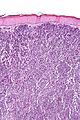
MCC - intermed. mag. (WC/Nephron)
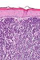
MCC - high mag. (WC/Nephron)
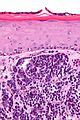
MCC - very high mag. (WC/Nephron)
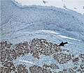
Merkel cell carcinoma - nested pattern (WC)


www:
- MCC (bccancer.bc.ca).
- MCC (joplink.net).
- Merkel cell carcinoma (ispub.com).
- Merkel cell carcinoma - several images (upmc.edu).
IHC
Features:
- CK7 -ve.
- CK20 +ve (perinuclear dot-like).[7]
- CAM5.2 +ve (dot-like pattern).
- CD56 +ve.
- AE1/AE3 +ve.
- Merkel cell polyomavirus +ve ~85% of cases.[8]
- NF +ve (12 of 13 cases).[9]
- Useful to differentiate from small cell carcinoma of lung.
Others:
EM
- Neurosecretory granules (AKA dense-core granules).[12]
See also
References
- ↑ 1.0 1.1 Calder, KB.; Smoller, BR. (May 2010). "New insights into merkel cell carcinoma.". Adv Anat Pathol 17 (3): 155-61. doi:10.1097/PAP.0b013e3181d97836. PMID 20418670.
- ↑ Pulitzer, MP.; Amin, BD.; Busam, KJ. (May 2009). "Merkel cell carcinoma: review.". Adv Anat Pathol 16 (3): 135-44. doi:10.1097/PAP.0b013e3181a12f5a. PMID 19395876.
- ↑ Feng, H.; Shuda, M.; Chang, Y.; Moore, PS. (Feb 2008). "Clonal integration of a polyomavirus in human Merkel cell carcinoma.". Science 319 (5866): 1096-100. doi:10.1126/science.1152586. PMID 18202256.
- ↑ Humphrey, Peter A; Dehner, Louis P; Pfeifer, John D (2008). The Washington Manual of Surgical Pathology (1st ed.). Lippincott Williams & Wilkins. pp. 491. ISBN 978-0781765275.
- ↑ 5.0 5.1 5.2 URL: /2011/SkinMerkelCell_11protocol.pdf http://www.cap.org/apps/docs/committees/cancer/cancer_protocols/2011/SkinMerkelCell_11protocol.pdf. Accessed on: 28 March 2012.
- ↑ 6.0 6.1 Ferringer, T.; Rogers, HC.; Metcalf, JS. (Feb 2005). "Merkel cell carcinoma in situ.". J Cutan Pathol 32 (2): 162-5. doi:10.1111/j.0303-6987.2005.00270.x. PMID 15606676.
- ↑ Leech, SN.; Kolar, AJ.; Barrett, PD.; Sinclair, SA.; Leonard, N. (Sep 2001). "Merkel cell carcinoma can be distinguished from metastatic small cell carcinoma using antibodies to cytokeratin 20 and thyroid transcription factor 1.". J Clin Pathol 54 (9): 727-9. PMID 11533085.
- ↑ Jung, HS.; Choi, YL.; Choi, JS.; Roh, JH.; Pyon, JK.; Woo, KJ.; Lee, EH.; Jang, KT. et al. (Oct 2011). "Detection of Merkel cell polyomavirus in Merkel cell carcinomas and small cell carcinomas by PCR and immunohistochemistry.". Histol Histopathol 26 (10): 1231-41. PMID 21870327.
- ↑ Bobos M, Hytiroglou P, Kostopoulos I, Karkavelas G, Papadimitriou CS (April 2006). "Immunohistochemical distinction between merkel cell carcinoma and small cell carcinoma of the lung". Am J Dermatopathol 28 (2): 99–104. doi:10.1097/01.dad.0000183701.67366.c7. PMID 16625069.
- ↑ Jankowski, M.; Kopinski, P.; Schwartz, R.; Czajkowski, R. (Nov 2014). "Merkel cell carcinoma: is this a true carcinoma?". Exp Dermatol 23 (11): 792-4. doi:10.1111/exd.12490. PMID 25040178.
- ↑ "Expression of anaplastic lymphoma kinase in Merkel cell carcinomas". Hum Pathol 44 (8): 1656–64. August 2013. doi:10.1016/j.humpath.2012.11.021. PMID 23574788.
- ↑ Gil-Moreno, A.; Garcia-Jiménez, A.; González-Bosquet, J.; Esteller, M.; Castellví-Vives, J.; Martínez Palones, JM.; Xercavins, J. (Mar 1997). "Merkel cell carcinoma of the vulva.". Gynecol Oncol 64 (3): 526-32. PMID 9062165.