Difference between revisions of "Mast cell"
Jump to navigation
Jump to search
| Line 30: | Line 30: | ||
*[[Neurofibroma]]. | *[[Neurofibroma]]. | ||
*[[Melanocytic nevi]].<ref name=pmid8432898>{{Cite journal | last1 = Carr | first1 = NJ. | last2 = Warren | first2 = AY. | title = Mast cell numbers in melanocytic naevi and cutaneous neurofibromas. | journal = J Clin Pathol | volume = 46 | issue = 1 | pages = 86-7 | month = Jan | year = 1993 | doi = | PMID = 8432898 }}</ref> | *[[Melanocytic nevi]].<ref name=pmid8432898>{{Cite journal | last1 = Carr | first1 = NJ. | last2 = Warren | first2 = AY. | title = Mast cell numbers in melanocytic naevi and cutaneous neurofibromas. | journal = J Clin Pathol | volume = 46 | issue = 1 | pages = 86-7 | month = Jan | year = 1993 | doi = | PMID = 8432898 }}</ref> | ||
*[[Succinate dehydrogenase-deficient renal cell carcinoma]]. | |||
==Stains== | ==Stains== | ||
Revision as of 06:15, 27 October 2014

The mast cell is an uncommonly cell that occasionally causes problems.
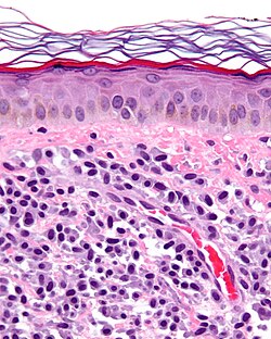
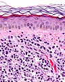
Microscopic
Features:
- Ovoid/round cell with moderate amount of gray granular cytoplasm.
- Nuclear ovoid/round.
- No obvious nucleolus.
- May be more abundant around blood vessels.
Notes:
- Lymphocyte vs. mast cell:
- Lymphocytes = round; mast cells = ovoid.
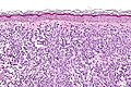
Images
www:
- Mastocytosis - low res. (jameswpattersonmd.com).
- Mastocytosis - bone marrow - several images (upmc.edu).
Mastocytosis - high mag. (WC)
Mastocytosis - very high mag. (WC)

Diseases
- Mastocytosis.
- Urticaria pigmentosa.
- Asthma.[1]
Conditions associated with the presence of mast cells
Stains
- Giemsa stain.
- Tyrosinase.[3] (???)
IHC
- CD117 +ve.
- CD34 +ve.[4][citation needed]
References
- ↑ Mitchell, Richard; Kumar, Vinay; Fausto, Nelson; Abbas, Abul K.; Aster, Jon (2011). Pocket Companion to Robbins & Cotran Pathologic Basis of Disease (8th ed.). Elsevier Saunders. pp. 370-2. ISBN 978-1416054542.
- ↑ Carr, NJ.; Warren, AY. (Jan 1993). "Mast cell numbers in melanocytic naevi and cutaneous neurofibromas.". J Clin Pathol 46 (1): 86-7. PMID 8432898.
- ↑ URL: http://www.nature.com/jid/journal/v53/n1/full/jid1969105a.html. Accessed on: 20 December 2011.
- ↑ Duşe, AO.; Ceauşu, RA.; Mezei, T.; Cîmpean, AM.; Gaje, P.; Ioniţă, H.; Jung, I. (2011). "Mast cells contribute to the angiogenesis in non-Hodgkin lymphoma. An immunohistochemical study based on the relationship with microvessel density.". Rom J Morphol Embryol 52 (3 Suppl): 1091-6. PMID 22119830.