Difference between revisions of "Lobular capillary hemangioma"
(redirect) |
|||
| (10 intermediate revisions by the same user not shown) | |||
| Line 1: | Line 1: | ||
{{ Infobox diagnosis | |||
| Name = {{PAGENAME}} | |||
| Image = SkinTumors-PB061062.JPG | |||
| Width = | |||
| Caption = Lobular capillary hemangioma. [[H&E stain]]. | |||
| Synonyms = pyogenic granuloma | |||
| Micro = polypoid ''or'' peduculated lesion, vascular - with plump endothelium, usu. thinned epithelium or ulcerated, lobular arrangement of vascular (seen at low power) | |||
| Subtypes = | |||
| LMDDx = [[capillary hemangioma]], [[myopericytoma]] (?), [[bacillary angiomatosis]], [[traumatic ulcerative granuloma with stromal eosinophilia]] - esp. for tongue lesions | |||
| Stains = | |||
| IHC = | |||
| EM = | |||
| Molecular = | |||
| IF = | |||
| Gross = | |||
| Grossing = | |||
| Site = [[Head and neck pathology|head and neck]] - lips, tongue, gingiva | |||
| Assdx = | |||
| Syndromes = | |||
| Clinicalhx = +/-rapid growth, young adults, children and pregnant women | |||
| Signs = | |||
| Symptoms = | |||
| Prevalence = | |||
| Bloodwork = | |||
| Rads = | |||
| Endoscopy = | |||
| Prognosis = benign | |||
| Other = | |||
| ClinDDx = | |||
| Tx = | |||
}} | |||
'''Lobular capillary hemangioma''', also known as '''pyogenic granuloma''',<ref name=pmid21839350>{{Cite journal | last1 = Baglin | first1 = AC. | title = [Vascular tumors and pseudotumors. Pyogenic granuloma (lobular capillary hemangioma)]. | journal = Ann Pathol | volume = 31 | issue = 4 | pages = 266-70 | month = Aug | year = 2011 | doi = 10.1016/j.annpat.2011.05.014 | PMID = 21839350 }}</ref> a benign head and neck lesion that can mimic malignancy. | |||
On occasion it is referred to as ''pregnancy tumour''. | |||
==General== | |||
*Seen in children, young adults, pregnant women. | |||
Clinical: | |||
*May grow quickly - clinically suspicious for a malignancy. | |||
Note: | |||
*''Pyogenic granuloma'' [[no truth in names|is a misnomer]]: | |||
**Not pyogenic, i.e. infectious. | |||
**Not [[granuloma|granulomatous]]. | |||
*The WMSP advocates the name ''lobular capillary hemangioma''.<ref name=Ref_WMSP12>{{Ref WMSP|12}}</ref> | |||
==Gross== | |||
Features:<ref name=Ref_PBoD776>{{Ref PBoD|776}}</ref> | |||
*Erythematous. | |||
*Hemorrhagic. | |||
Usually location:<ref name=Ref_WMSP12>{{Ref WMSP|12}}</ref> | |||
*Lips. | |||
*[[Tongue]]. | |||
*Gingiva. | |||
==Microscopic== | |||
Features:<ref name=Ref_PBoD775>{{Ref PBoD|775}}</ref> | |||
*Polypoid ''or'' peduculated. | |||
*Vascular, i.e. many blood vessels, with plump endothelium. | |||
*Usu. thinned epithelium<ref>URL: [http://basicpathology-histopathology.blogspot.com/2009/10/head-and-neck-oral-cavity-reactive_3282.html http://basicpathology-histopathology.blogspot.com/2009/10/head-and-neck-oral-cavity-reactive_3282.html]. Accessed on: 2 February 2011.</ref> or ulcerated.<ref name=Ref_WMSP12>{{Ref WMSP|12}}</ref> | |||
*Lobular arrangement of vascular (seen at low power).<ref>S. Sade. 8 September 2011.</ref> | |||
DDx: | |||
*[[Capillary hemangioma]]. | |||
*[[Myopericytoma]] (???). | |||
*[[Bacillary angiomatosis]].<ref name=pmid16310070>{{Cite journal | last1 = Levy | first1 = I. | last2 = Rolain | first2 = JM. | last3 = Lepidi | first3 = H. | last4 = Raoult | first4 = D. | last5 = Feinmesser | first5 = M. | last6 = Lapidoth | first6 = M. | last7 = Ben-Amitai | first7 = D. | title = Is pyogenic granuloma associated with Bartonella infection? | journal = J Am Acad Dermatol | volume = 53 | issue = 6 | pages = 1065-6 | month = Dec | year = 2005 | doi = 10.1016/j.jaad.2005.08.046 | PMID = 16310070 }}</ref> | |||
*[[Traumatic ulcerative granuloma with stromal eosinophilia]] - abundant [[eosinophil]]s. | |||
Why it is not... | |||
*[[Glomus tumour]] - cookie cutter arrangement of cells. | |||
===Image=== | |||
<gallery> | |||
Image:SkinTumors-PB061062.JPG | Pyogenic granuloma. (WC) | |||
</gallery> | |||
www: | |||
*[http://www.sciencephoto.com/images/download_lo_res.html?id=670066054 Pyogenic granuloma (sciencephoto.com)]. | |||
==IHC== | |||
Features - positive for vascular markers:<ref name=Ref_WMSP12>{{Ref WMSP|12}}</ref> | |||
*CD34 +ve. | |||
*CD31 +ve. | |||
*Factor VIII +ve. | |||
*SMA +ve -- marks pericytes.{{fact}} | |||
A panel: | |||
*CD31, SMA, HMB-45. | |||
==Sign out== | |||
<pre> | |||
Tongue Lesion, Excision: | |||
- Lobular capillary hemangioma (pyogenic granuloma). | |||
</pre> | |||
===Block letters=== | |||
<pre> | |||
TONGUE, LEFT LATERAL, BIOPSY: | |||
- LOBULAR CAPILLARY HEMANGIOMA (PYOGENIC GRANULOMA). | |||
</pre> | |||
<pre> | |||
LESION, POSTERIOR TO LEFT EAR, EXCISION: | |||
- LOBULAR CAPILLARY HEMANGIOMA (PYOGENIC GRANULOMA). | |||
COMMENT: | |||
The lesion stains as follows: | |||
POSITIVE: SMA, CD31. | |||
NEGATIVE: S-100, HMB-45. | |||
</pre> | |||
===Micro=== | |||
The sections shows a pendunculated vascular lesion with small capillaries arranged in a lobular fashion. The endothelial cells of the lesion show no atypia. The overlying acanthotic epidermis has hyperkeratosis and hypergranulosis, and is focally ulcerated and impetiginized. There is no significant keratocyte atypia. No melanocytic nests are seen. The dermis has a mild perivascular lymphoplasmacytic infiltrate. The lesion is excised in the plane of section. | |||
====Alternate - tongue==== | |||
The sections show a pendunculated vascular lesion with small capillaries arranged in a lobular fashion. The endothelial cells of the lesion show no significant atypia. The overlying epithelium is largely eroded. There is no significant keratocyte atypia. The lesion is excised in the plane of section. | |||
==See also== | |||
*[[Head and neck pathology]]. | |||
==References== | |||
{{Reflist|2}} | |||
[[Category:Diagnosis]] | |||
[[Category:Head and neck pathology]] | |||
Latest revision as of 18:52, 18 January 2022
| Lobular capillary hemangioma | |
|---|---|
| Diagnosis in short | |
 Lobular capillary hemangioma. H&E stain. | |
|
| |
| Synonyms | pyogenic granuloma |
|
| |
| LM | polypoid or peduculated lesion, vascular - with plump endothelium, usu. thinned epithelium or ulcerated, lobular arrangement of vascular (seen at low power) |
| LM DDx | capillary hemangioma, myopericytoma (?), bacillary angiomatosis, traumatic ulcerative granuloma with stromal eosinophilia - esp. for tongue lesions |
| Site | head and neck - lips, tongue, gingiva |
|
| |
| Clinical history | +/-rapid growth, young adults, children and pregnant women |
| Prognosis | benign |
Lobular capillary hemangioma, also known as pyogenic granuloma,[1] a benign head and neck lesion that can mimic malignancy.
On occasion it is referred to as pregnancy tumour.
General
- Seen in children, young adults, pregnant women.
Clinical:
- May grow quickly - clinically suspicious for a malignancy.
Note:
- Pyogenic granuloma is a misnomer:
- Not pyogenic, i.e. infectious.
- Not granulomatous.
- The WMSP advocates the name lobular capillary hemangioma.[2]
Gross
Features:[3]
- Erythematous.
- Hemorrhagic.
Usually location:[2]
- Lips.
- Tongue.
- Gingiva.
Microscopic
Features:[4]
- Polypoid or peduculated.
- Vascular, i.e. many blood vessels, with plump endothelium.
- Usu. thinned epithelium[5] or ulcerated.[2]
- Lobular arrangement of vascular (seen at low power).[6]
DDx:
- Capillary hemangioma.
- Myopericytoma (???).
- Bacillary angiomatosis.[7]
- Traumatic ulcerative granuloma with stromal eosinophilia - abundant eosinophils.
Why it is not...
- Glomus tumour - cookie cutter arrangement of cells.
Image
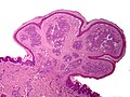
Pyogenic granuloma. (WC)
www:
IHC
Features - positive for vascular markers:[2]
- CD34 +ve.
- CD31 +ve.
- Factor VIII +ve.
- SMA +ve -- marks pericytes.[citation needed]
A panel:
- CD31, SMA, HMB-45.
Sign out
Tongue Lesion, Excision:
- Lobular capillary hemangioma (pyogenic granuloma).
Block letters
TONGUE, LEFT LATERAL, BIOPSY:
- LOBULAR CAPILLARY HEMANGIOMA (PYOGENIC GRANULOMA).
LESION, POSTERIOR TO LEFT EAR, EXCISION:
- LOBULAR CAPILLARY HEMANGIOMA (PYOGENIC GRANULOMA).

COMMENT:
The lesion stains as follows:
POSITIVE: SMA, CD31.
NEGATIVE: S-100, HMB-45.
Micro
The sections shows a pendunculated vascular lesion with small capillaries arranged in a lobular fashion. The endothelial cells of the lesion show no atypia. The overlying acanthotic epidermis has hyperkeratosis and hypergranulosis, and is focally ulcerated and impetiginized. There is no significant keratocyte atypia. No melanocytic nests are seen. The dermis has a mild perivascular lymphoplasmacytic infiltrate. The lesion is excised in the plane of section.
Alternate - tongue
The sections show a pendunculated vascular lesion with small capillaries arranged in a lobular fashion. The endothelial cells of the lesion show no significant atypia. The overlying epithelium is largely eroded. There is no significant keratocyte atypia. The lesion is excised in the plane of section.
See also
References
- ↑ Baglin, AC. (Aug 2011). "[Vascular tumors and pseudotumors. Pyogenic granuloma (lobular capillary hemangioma)].". Ann Pathol 31 (4): 266-70. doi:10.1016/j.annpat.2011.05.014. PMID 21839350.
- ↑ 2.0 2.1 2.2 2.3 Humphrey, Peter A; Dehner, Louis P; Pfeifer, John D (2008). The Washington Manual of Surgical Pathology (1st ed.). Lippincott Williams & Wilkins. pp. 12. ISBN 978-0781765275.
- ↑ Cotran, Ramzi S.; Kumar, Vinay; Fausto, Nelson; Nelso Fausto; Robbins, Stanley L.; Abbas, Abul K. (2005). Robbins and Cotran pathologic basis of disease (7th ed.). St. Louis, Mo: Elsevier Saunders. pp. 776. ISBN 0-7216-0187-1.
- ↑ Cotran, Ramzi S.; Kumar, Vinay; Fausto, Nelson; Nelso Fausto; Robbins, Stanley L.; Abbas, Abul K. (2005). Robbins and Cotran pathologic basis of disease (7th ed.). St. Louis, Mo: Elsevier Saunders. pp. 775. ISBN 0-7216-0187-1.
- ↑ URL: http://basicpathology-histopathology.blogspot.com/2009/10/head-and-neck-oral-cavity-reactive_3282.html. Accessed on: 2 February 2011.
- ↑ S. Sade. 8 September 2011.
- ↑ Levy, I.; Rolain, JM.; Lepidi, H.; Raoult, D.; Feinmesser, M.; Lapidoth, M.; Ben-Amitai, D. (Dec 2005). "Is pyogenic granuloma associated with Bartonella infection?". J Am Acad Dermatol 53 (6): 1065-6. doi:10.1016/j.jaad.2005.08.046. PMID 16310070.