Difference between revisions of "Intranodal palisaded myofibroblastoma"
Jump to navigation
Jump to search
(more) |
m (another image) |
||
| (29 intermediate revisions by 2 users not shown) | |||
| Line 1: | Line 1: | ||
{{ Infobox diagnosis | |||
| Name = {{PAGENAME}} | |||
| Image = Intranodal palisaded myofibroblastoma - very high mag.jpg | |||
| Width = 200 | |||
| Caption = IPM. [[H&E stain]]. | |||
| Micro = spindle cells with nuclear palisading, RBC extravasation | |||
| Subtypes = | |||
| LMDDx = [[schwannoma]], [[leiomyoma]] | |||
| Stains = | |||
| IHC = SMA +ve, S-100 -ve. | |||
| EM = | |||
| Molecular = | |||
| IF = | |||
| Gross = | |||
| Grossing = | |||
| Site = [[lymph node]]. | |||
| Assdx = | |||
| Syndromes = | |||
| Clinicalhx = | |||
| Signs = mass lesion | |||
| Symptoms = | |||
| Prevalence = very rare. | |||
| Bloodwork = | |||
| Rads = | |||
| Endoscopy = | |||
| Prognosis = | |||
| Other = | |||
| ClinDDx = [[lymphoma]], metastatic carcinoma. | |||
}} | |||
'''Intranodal palisaded myofibroblastoma''', abbreviated '''IPM''', is a rare tumour that classically presents as an inguinal mass.<ref name=pmid17284119>{{Cite journal | last1 = Nguyen | first1 = T. | last2 = Eltorky | first2 = MA. | title = Intranodal palisaded myofibroblastoma. | journal = Arch Pathol Lab Med | volume = 131 | issue = 2 | pages = 306-10 | month = Feb | year = 2007 | doi = 10.1043/1543-2165(2007)131[306:IPM]2.0.CO;2 | PMID = 17284119 }}</ref> | '''Intranodal palisaded myofibroblastoma''', abbreviated '''IPM''', is a rare tumour that classically presents as an inguinal mass.<ref name=pmid17284119>{{Cite journal | last1 = Nguyen | first1 = T. | last2 = Eltorky | first2 = MA. | title = Intranodal palisaded myofibroblastoma. | journal = Arch Pathol Lab Med | volume = 131 | issue = 2 | pages = 306-10 | month = Feb | year = 2007 | doi = 10.1043/1543-2165(2007)131[306:IPM]2.0.CO;2 | PMID = 17284119 }}</ref> | ||
==General== | ==General== | ||
*Rare. | *Rare ~ 55 cases in the world literature.<ref name=pmid21718465/> | ||
Demographics: | |||
*Male:female = 2:1. | *Male:female = 2:1. | ||
*Adults - middle age. | *Adults - middle age. | ||
Location: | |||
*Usually inguinal lymph node. | |||
*Reported in retroperitoneum.<ref name=pmid21718465>{{Cite journal | last1 = Sagar | first1 = J. | last2 = Vargiamidou | first2 = A. | last3 = Manikkapurath | first3 = H. | title = Intranodal palisaded myofibroblastoma originating from retroperitoneum: an unusual origin. | journal = BMC Clin Pathol | volume = 11 | issue = | pages = 7 | month = | year = 2011 | doi = 10.1186/1472-6890-11-7 | PMID = 21718465 | PMC = 3146916 }}</ref> | |||
Treatment: | |||
*Simple excision; rare recurrences have been reported.<ref name=pmid10235504>{{Cite journal | last1 = Creager | first1 = AJ. | last2 = Garwacki | first2 = CP. | title = Recurrent intranodal palisaded myofibroblastoma with metaplastic bone formation. | journal = Arch Pathol Lab Med | volume = 123 | issue = 5 | pages = 433-6 | month = May | year = 1999 | doi = 10.1043/0003-9985(1999)1230433:RIPMWM2.0.CO;2 | PMID = 10235504 }}</ref> | |||
==Microscopic== | ==Microscopic== | ||
Features: | Features: | ||
#Rim of peripheral lymphoid tissue. | #Rim of peripheral lymphoid tissue. | ||
# | #*Remnant of [[lymph node]]. | ||
#RBC extravasation/hemorrhage. | #Spindle cells with nuclear palisading - '''key feature'''. | ||
#Amianthoid fibers - blood vessel surrounded by collagen with peripheral spokes.<ref name=pmid1918406>{{Cite journal | last1 = Bigotti | first1 = G. | last2 = Coli | first2 = A. | last3 = Mottolese | first3 = M. | last4 = Di Filippo | first4 = F. | title = Selective location of palisaded myofibroblastoma with amianthoid fibres. | journal = J Clin Pathol | volume = 44 | issue = 9 | pages = 761-4 | month = Sep | year = 1991 | doi = | PMID = 1918406 }}</ref> | #[[RBC extravasation]]/hemorrhage. | ||
# | #Amianthoid fibers - blood vessel surrounded by collagen with (fine) peripheral spokes.<ref name=pmid1918406>{{Cite journal | last1 = Bigotti | first1 = G. | last2 = Coli | first2 = A. | last3 = Mottolese | first3 = M. | last4 = Di Filippo | first4 = F. | title = Selective location of palisaded myofibroblastoma with amianthoid fibres. | journal = J Clin Pathol | volume = 44 | issue = 9 | pages = 761-4 | month = Sep | year = 1991 | doi = | PMID = 1918406 }}</ref> | ||
#*Paucicellular regions.<ref>URL: [http://path.upmc.edu/cases/case121/micro.html http://path.upmc.edu/cases/case121/micro.html]. Accessed on: 3 January 2012.</ref> | |||
#Intracellular and extracellular fuchsinophilic bodies. | |||
#*Smooth muscle actin +ve. | |||
Notes: | |||
*''Fuchsinophilic'' = affinity for the acid dye fuchsin.<ref>URL: [http://www.merriam-webster.com/medical/fuchsinophilic http://www.merriam-webster.com/medical/fuchsinophilic]. Accessed on: 3 October 2011.</ref> | |||
**Image: [http://www.flickr.com/photos/53376324@N08/5387821397/in/photostream Fuchsinophilic material (flickr.com)] - red. | |||
DDx: | DDx:<ref>{{Cite journal | last1 = Sarma | first1 = NH. | last2 = Arora | first2 = KS. | last3 = Varalaxmi | first3 = KP. | title = Intranodal palisaded myofibroblastoma: a case report and an update on etiopathogenesis and differential diagnosis. | journal = J Cancer Res Ther | volume = 9 | issue = 2 | pages = 295-8 | month = | year = | doi = 10.4103/0973-1482.113395 | PMID = 23771380 }}</ref> | ||
*[[Schwannoma]]. | *[[Schwannoma]]. | ||
*[[Leiomyoma]]. | |||
*[[Leiomyosarcoma]]. | |||
*[[Malignant melanoma]]. | |||
*[[Kaposi sarcoma]]. | |||
*Metastatic carcinoma. | |||
Images: | ===Images=== | ||
<gallery> | |||
Image: Intranodal palisaded myofibroblastoma - low mag.jpg | IPM - low mag. (WC/Nephron) | |||
Image: Intranodal palisaded myofibroblastoma - intermed mag.jpg | IPM - intermed. mag. (WC/Nephron) | |||
Image: Intranodal palisaded myofibroblastoma - high mag.jpg | IPM - high mag. (WC/Nephron) | |||
Image: Intranodal palisaded myofibroblastoma - very high mag.jpg | IPM - very high mag. (WC/Nephron) | |||
Image: Intra-nodal palisaded myofibroblastoma.jpg | IPM - high mag. (WC) | |||
</gallery> | |||
www: | |||
*[http://www.surgicalpathologyatlas.com/glfusion/mediagallery/media.php?f=0&sort=0&s=20080802163636511 IPM (surgicalpathologyatlas.com)]. | *[http://www.surgicalpathologyatlas.com/glfusion/mediagallery/media.php?f=0&sort=0&s=20080802163636511 IPM (surgicalpathologyatlas.com)]. | ||
*[http://www.ncbi.nlm.nih.gov/pmc/articles/PMC496726/figure/F4/ Amianthoid fibers (nih.gov)].<ref name=pmid1918406>{{Cite journal | last1 = Bigotti | first1 = G. | last2 = Coli | first2 = A. | last3 = Mottolese | first3 = M. | last4 = Di Filippo | first4 = F. | title = Selective location of palisaded myofibroblastoma with amianthoid fibres. | journal = J Clin Pathol | volume = 44 | issue = 9 | pages = 761-4 | month = Sep | year = 1991 | doi = | PMID = 1918406 }}</ref> | *[http://www.ncbi.nlm.nih.gov/pmc/articles/PMC496726/figure/F4/ Amianthoid fibers (nih.gov)].<ref name=pmid1918406>{{Cite journal | last1 = Bigotti | first1 = G. | last2 = Coli | first2 = A. | last3 = Mottolese | first3 = M. | last4 = Di Filippo | first4 = F. | title = Selective location of palisaded myofibroblastoma with amianthoid fibres. | journal = J Clin Pathol | volume = 44 | issue = 9 | pages = 761-4 | month = Sep | year = 1991 | doi = | PMID = 1918406 }}</ref> | ||
*[http://www.ncbi.nlm.nih.gov/pmc/articles/PMC3146916/figure/F4/ Amianthoid fibers (nih.gov)].<ref name=pmid21718465/> | |||
*[http://path.upmc.edu/cases/case121/micro.html IPM - several images (upmc.edu)]. | |||
==IHC== | ==IHC== | ||
*SMA +ve. | *SMA +ve. | ||
*Cyclin D1 +ve. | *Vimentin +ve. | ||
*Cyclin D1 +ve/-ve.<ref name=pmid23771380>{{Cite journal | last1 = Sarma | first1 = NH. | last2 = Arora | first2 = KS. | last3 = Varalaxmi | first3 = KP. | title = Intranodal palisaded myofibroblastoma: a case report and an update on etiopathogenesis and differential diagnosis. | journal = J Cancer Res Ther | volume = 9 | issue = 2 | pages = 295-8 | month = | year = | doi = 10.4103/0973-1482.113395 | PMID = 23771380 }}</ref> | |||
Other: | Other: | ||
| Line 32: | Line 93: | ||
*Desmin -ve | *Desmin -ve | ||
*Ki-67 - low. | *Ki-67 - low. | ||
*HMB-45 -ve. | |||
==See also== | ==See also== | ||
| Line 41: | Line 103: | ||
[[Category:Lymph node pathology]] | [[Category:Lymph node pathology]] | ||
[[Category:Diagnosis]] | |||
Latest revision as of 16:54, 23 September 2018
| Intranodal palisaded myofibroblastoma | |
|---|---|
| Diagnosis in short | |
 IPM. H&E stain. | |
|
| |
| LM | spindle cells with nuclear palisading, RBC extravasation |
| LM DDx | schwannoma, leiomyoma |
| IHC | SMA +ve, S-100 -ve. |
| Site | lymph node. |
|
| |
| Signs | mass lesion |
| Prevalence | very rare. |
| Clin. DDx | lymphoma, metastatic carcinoma. |
Intranodal palisaded myofibroblastoma, abbreviated IPM, is a rare tumour that classically presents as an inguinal mass.[1]
General
- Rare ~ 55 cases in the world literature.[2]
Demographics:
- Male:female = 2:1.
- Adults - middle age.
Location:
- Usually inguinal lymph node.
- Reported in retroperitoneum.[2]
Treatment:
- Simple excision; rare recurrences have been reported.[3]
Microscopic
Features:
- Rim of peripheral lymphoid tissue.
- Remnant of lymph node.
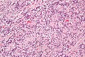
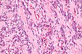
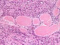
- Spindle cells with nuclear palisading - key feature.
- RBC extravasation/hemorrhage.
- Amianthoid fibers - blood vessel surrounded by collagen with (fine) peripheral spokes.[4]
- Paucicellular regions.[5]
- Intracellular and extracellular fuchsinophilic bodies.
- Smooth muscle actin +ve.
Notes:
- Fuchsinophilic = affinity for the acid dye fuchsin.[6]
- Image: Fuchsinophilic material (flickr.com) - red.
DDx:[7]
- Schwannoma.
- Leiomyoma.
- Leiomyosarcoma.
- Malignant melanoma.
- Kaposi sarcoma.
- Metastatic carcinoma.
Images
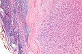
IPM - low mag. (WC/Nephron)

IPM - intermed. mag. (WC/Nephron)
IPM - high mag. (WC/Nephron)
IPM - very high mag. (WC/Nephron)
IPM - high mag. (WC)


www:
- IPM (surgicalpathologyatlas.com).
- Amianthoid fibers (nih.gov).[4]
- Amianthoid fibers (nih.gov).[2]
- IPM - several images (upmc.edu).
IHC
- SMA +ve.
- Vimentin +ve.
- Cyclin D1 +ve/-ve.[8]
Other:
- S100 -ve
- Excludes schwannoma.
- GFAP -ve.
- CD34 -ve.
- Desmin -ve
- Ki-67 - low.
- HMB-45 -ve.
See also
References
- ↑ Nguyen, T.; Eltorky, MA. (Feb 2007). "Intranodal palisaded myofibroblastoma.". Arch Pathol Lab Med 131 (2): 306-10. doi:10.1043/1543-2165(2007)131[306:IPM]2.0.CO;2. PMID 17284119.
- ↑ 2.0 2.1 2.2 Sagar, J.; Vargiamidou, A.; Manikkapurath, H. (2011). "Intranodal palisaded myofibroblastoma originating from retroperitoneum: an unusual origin.". BMC Clin Pathol 11: 7. doi:10.1186/1472-6890-11-7. PMC 3146916. PMID 21718465. https://www.ncbi.nlm.nih.gov/pmc/articles/PMC3146916/.
- ↑ Creager, AJ.; Garwacki, CP. (May 1999). "Recurrent intranodal palisaded myofibroblastoma with metaplastic bone formation.". Arch Pathol Lab Med 123 (5): 433-6. doi:10.1043/0003-9985(1999)1230433:RIPMWM2.0.CO;2. PMID 10235504.
- ↑ 4.0 4.1 Bigotti, G.; Coli, A.; Mottolese, M.; Di Filippo, F. (Sep 1991). "Selective location of palisaded myofibroblastoma with amianthoid fibres.". J Clin Pathol 44 (9): 761-4. PMID 1918406.
- ↑ URL: http://path.upmc.edu/cases/case121/micro.html. Accessed on: 3 January 2012.
- ↑ URL: http://www.merriam-webster.com/medical/fuchsinophilic. Accessed on: 3 October 2011.
- ↑ Sarma, NH.; Arora, KS.; Varalaxmi, KP.. "Intranodal palisaded myofibroblastoma: a case report and an update on etiopathogenesis and differential diagnosis.". J Cancer Res Ther 9 (2): 295-8. doi:10.4103/0973-1482.113395. PMID 23771380.
- ↑ Sarma, NH.; Arora, KS.; Varalaxmi, KP.. "Intranodal palisaded myofibroblastoma: a case report and an update on etiopathogenesis and differential diagnosis.". J Cancer Res Ther 9 (2): 295-8. doi:10.4103/0973-1482.113395. PMID 23771380.