Intradermal nevus
Jump to navigation
Jump to search
| Intradermal nevus | |
|---|---|
| Diagnosis in short | |
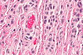
 Intradermal nevus. H&E stain. | |
|
| |
| Synonyms | intradermal melanocytic nevus |
|
| |
| LM | nests of melanocytes in dermis (only), melanocytes "mature" with depth, usu. no mitoses (occ. superficial), no destruction of surrounding structures, no conspicuous nucleoli, no significant melanocyte enlargement |
| LM DDx | malignant melanoma (nevoid), junctional nevus, compound nevus, dysplastic nevus, skin tag |
| Gross | pigment skin lesion, usu. small, regular border, no irregularity in pigmentation |
| Site | skin - see melanocytic lesions and common nevus |
|
| |
| Prevalence | very common |
| Prognosis | benign |
| Clin. DDx | pigmented skin lesions |
| Treatment | none required, may be excised for cosmetic reasons |
Intradermal nevus (abbreviated IDN), also intradermal melanocytic nevus, is a common benign melanocytic lesion.
The intradermal nevus is in the large group common nevus. In common language, nevus is known as a mole.
General
- Benign.
- Common.
- Think melanoma.
Clinical:
- ABCD = asymmetric, borders (irregular), colour (black), diameter (large).
Microscopic
Features:
- Symmetrical lesion.
- "Matures" with depth.
- Less cellular with depth.
- Less nuclear atypia with depth.
- Smaller cells with depth.
- Smaller nests with depth.
- Rare mitoses (superficial).
- No deep mitoses.
- No destruction of surrounding structures.
- No nucleoli.
- In the dermis only - key feature.
- +/-Adipocytes - uncommon.[1]
DDx:
- Malignant melanoma (nevoid).
- Dysplastic nevus.
- Junctional nevus.
- Compound nevus.
- Skin tag.
Images
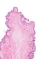
IDN - very low mag.
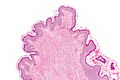
IDN - low mag.
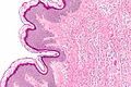
IDN - intermed. mag.
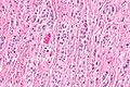
IDN - high mag.
IDN - very high mag.


