Difference between revisions of "Gross pathology"

From Libre Pathology
Jump to navigation Jump to search
m (+image)
Line 24: Line 24:
*[[Calcific aortic stenosis]].


==Gross spot diagnosis==
==Gross pathology spot diagnoses==
===Heart===
{{Main|Gross pathology spot diagnoses}}
[[Heart valve]]:
*[[Infective endocarditis]].
*[[Bicuspid aortic valve]].
*[[Nonbacterial thrombotic endocarditis]] - non-distructive.
*[[Rheumatic heart disease]] - fish mouth valve, thick cordae tendinae.
*[[Calcific aortic stenosis]].
Heart - other:
*[[Fibrinous pericarditis]].
*[[Hypertrophic cardiomyopathy]] - septal thickness > free wall thickness.
*Cardiac hypertrophy.
*[[Dilated cardiomyopathy]].
*[[Arrhythmogenic right ventricular cardiomyopathy]].
*[[Atrial septal defect]].
*[[Ventricular septal defect]].
*Patent foramen ovale.
 
===Cardiovascular===
*[[Aortic dissection]].
*Aortic rupture.
*[[Cerebral saccular aneurysm]].
 
===Lungs===
*[[Pulmonary thromboembolism]].
*[[Pulmonary infarct]].
*Lung abscess.
*Lung tumour (primary).
*Metatstatic tumour lung - usu. multiple and peripheral.
*[[Malignant mesothelioma]].
*[[Pleural plaques]].
*[[Emphysema]].
*[[Pneumothorax]].
*[[Hemothorax]].
*Hypoplasia secondary to [[diaphragmatic hernia]].
 
===Gastrointestinal pathology===
[[Esophagus]]:
*[[Squamous cell carcinoma of the esophagus]] - classically mid esophagus.
*[[Adenocarcinoma of the esophagus]] - usually distal esophagus.
*[[Barrett's esophagus]].
*[[Esophageal varices]].
*Esophageal perforation.
 
Stomach:
*Malignant gastric ulcer.
*Peptic ulcer.
*[[Gastric antral vascular ectasia]].
*[[Leopard spots]] - seen in hypothermia.
*[[GIST]].
 
[[Duodenum]]:
*Peptic ulcer.
*[[Duodenal adenocarcinoma]] - classically assoc. with [[FAP]].
 
[[Liver]]:
*[[Cirrhosis]].
*[[Hydatid cyst]].
*[[Hepatocellular carcinoma]] - in the context of cirrhosis.
*[[Metastases]].
*[[Hemangioma]].
*[[Focal nodular hyperplasia]].
*[[Hepatic adenoma]].
*[[Cholangiocarcinoma]].
*[[Congestive hepatopathy]] (nutmeg liver) - seen in [[congestive heart failure]].
*Hepatic infarct - classically at the periphery, red and wedge-shaped.
 
[[Gallbladder]]:
*[[Cholelithiasis]].
*[[Cholesterolosis of the gallbladder]].
*Gallbladder polyp.
 
[[Pancreas]]:
*[[Solid pseudopapillary neoplasm]] - with age & sex provided.
*[[Pancreatic adenocarcinoma]].
*[[Neuroendocrine tumour]].
*[[Chronic pancreatitis]].
 
[[Small bowel]]:
*Ischemic small bowel.
*[[Meckel's diverticulum]].
*[[Intussusception]].
*Neuroendocrine tumour - if on section.
*[[GIST]].
*Incarcerated hernia.
 
[[Appendix]]:
*[[Acute appendicitis]].
*[[Neuroendocrine tumour of the appendix]].
*[[Mucinous tumour of the appendix]].
 
Large bowel:
*[[Colorectal adenocarcinoma]].
*Bowel infarction.
*[[Ulcerative colitis]] - no skip lesions, no wall thickening.
*[[Crohn's disease]] - [[creeping fat]], cobble stone pattern, skip lesions, fistulas.
*[[Pseudomembranous colitis]].
 
===Gynecologic pathology===
Ovary:
*Mucinous tumour.
*[[Mature teratoma]].
*[[Serous cystadenoma of the ovary]].
*[[ovarian fibroma|Fibroma]].
*[[Brenner tumour]] - may be yellow, typically solid.
*[[Endometrioma]].
 
Uterine tube:
*[[Ectopic pregnancy]].
*Tubal abscess.
*[[Adenomatoid tumour]].
 
Uterus:
*[[Adenomyosis of the uterus|Adenomyosis]].
*[[Uterine leiomyoma|Leiomyoma]] - multiple.
*Endometrial carcinoma.
*[[Uterine leiomyosarcoma|Leiomyosarcoma]] - friable, solitary, fish flesh.
*[[Bicornuate uterus]].
*[[Uterus didelphys]].
*[[Sarcoma botryoides]] (embryonal rhabdomyosarcoma).
 
Uterine cervix:
*[[Nabothian cyst]].
*[[Cervical polyp]].
*Cervical carcinoma.
 
===Dermatopathology===
Tumour:
*[[Basal cell carcinoma]].
*[[Squamous cell carcinoma]].
*[[Malignant melanoma]].
 
Possible syndromic:
*[[Seborrheic keratosis]] - [[Leser–Trélat sign]]
*[[Neurofibroma]] - [[neurofibromatosis]].
*[[Angiofibroma]] - [[Fabry disease]].
 
===Neuropathology===
Bleeds:
*[[Epidural hematoma]]
*[[Subdural hematoma]].
*[[Subarachnoid hematoma]] - [[berry aneurysm]].
*[[Intracerebral hematoma]] - hypertensive, usu. thalamus.
**Lobar hemorrhage - periphery of cortex.
*[[Duret hemorrhage]] - pontine hemorrhage.
 
Tumours:
*[[Meningioma]] - intradural, extramedullary.
*Glioma.
*[[Schwannoma]] - cerebellopontine angle.
*[[Myxopapillary ependymoma]] - filum terminale.
 
Non-tumour:
*[[Diffuse axonal injury]] - classically corpus callosum.
*[[Fat embolism]].
*[[Cerebral contusion]].
**[[Plaque jaune]].
*Cerebral infarction - classically MCA territory.
 
Neurodegenerative:
*[[Alzheimer disease]] - atrophy spares occipital lobe.
*Frontotemporal dementia.
*[[Huntington disease]] - young, caudate nucleus missing.
 
===Genitourinary pathology===
Kidney:
*[[Clear cell renal cell carcinoma]] - yellow.
*[[Papillary renal cell carcinoma]] - friable.
*[[Chromophobe renal cell carcinoma]] - brown +/-hemorrhage, no central scar.
*[[Renal oncocytoma]] - brown, central scar.
*[[Autosomal dominant polycystic kidney disease]].
*[[Angiomyolipoma]] - fat.
 
Testis:
*[[Seminoma]] - solid.
*[[Mixed germ cell tumour]] - cystic (often teratoma) & solid.<ref name=Ref_AoGP447>{{Ref AoGP|447}}</ref>
*[[Leydig cell tumour]] - brown.
*Testicular torsion.
 
Paratesticular:
*[[Liposarcoma]].
 
===Placenta===
Membranes:
*Marginal inserion - normal.
*[[Circumvallate placenta]].
*[[Circummarginate placenta]].
 
Disc:
*Retroplacental blood clot - suggestive of [[placental abruption]].
*[[Chorangioma]] - big red intraparenchymal.
*[[Placental infarct]] - white = old, red = recent.
*[[Twin-to-twin transfusion syndrome]] - one side beefy red the other pale.
*Meconium staining/[[chorioamnionitis]].
*[[Amnion nodosum]].
*[[Squamous metaplasia of the amnion]].
*Succenturiate placenta (placenta with accessory lobe(s)).
*Bilobate placenta.
 
Cord:
*[[Two vessel umbilical cord]].
*False knot.
*True knot.
*Membranous insertion ([[AKA]] velamentous insertion).
*Furcate insertion - vessels separate before inserting.
 
===Fetal===
*[[Amniotic rupture sequence]] - includes [[amniotic band syndrome]].
*[[Twin-to-twin transfusion syndrome]] (TTTS).
 
Genetic:
*[[Down syndrome]].
*[[Edwards syndrome]].
*[[Patau syndrome]].
*[[Turner syndrome]].


==See also==
==See also==

Revision as of 17:22, 27 July 2014

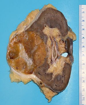
A kidney tumour (renal oncocytoma) at the time of grossing.

Gross pathology, also simply gross, refers to the macroscopic pathology, and the macroscopic assessment of pathology specimens. It may include preparation of tissue for a microscopic examination. It is an essential part of pathologic assessments.

The process of cutting up specimens is known as grossing (North American term), cut-up or macroscopic cut-up (Australian term).

Specimen opening

Usually referred to simply as opening.
May go by the term freshing.
  • The first part of a gross pathologic assessment.

Components

  1. Orient the specimen.
  2. Paint with ink - if applicable.
    • A good general rule is: ink before you think.
  3. Cut open for fixation - if not immediately blocked.

Gross only

Which specimens are "gross only" typically depends on institutional policy.[1]

Common gross only specimens

Gross pathology spot diagnoses

See also

References

  1. Zarbo, RJ.; Nakhleh, RE. (Feb 1999). "Surgical pathology specimens for gross examination only and exempt from submission: a College of American Pathologists Q-Probes study of current policies in 413 institutions.". Arch Pathol Lab Med 123 (2): 133-9. doi:10.1043/0003-9985(1999)1230133:SPSFGE2.0.CO;2. PMID 10050786.

External links