Difference between revisions of "Granulocytic sarcoma"
(+cat.) |
m (cases can occasionally precede full blown AML) |
||
| (11 intermediate revisions by 2 users not shown) | |||
| Line 1: | Line 1: | ||
{{ Infobox diagnosis | |||
| Name = {{PAGENAME}} | |||
| Image = Chloroma_-_very_high_mag.jpg | |||
| Width = | |||
| Caption = Chloroma. [[H&E stain]]. | |||
| Synonyms = extramedullary leukemia, myeloid sarcoma and chloroma, myeloblastoma, chloromyeloma, chloromyelosarcoma, granulocytic leukosarcoma, myelosarcoma | |||
| Micro = atypical small blue cells ~2x resting lymphocyte, infiltrative | |||
| Subtypes = | |||
| LMDDx = [[small round cell tumours]] | |||
| Stains = | |||
| IHC = CD117 +ve, CD43 +ve, CD34 +ve/-ve | |||
| EM = | |||
| Molecular = | |||
| IF = | |||
| Gross = | |||
| Grossing = | |||
| Site = [[soft tissue lesion]] | |||
| Assdx = | |||
| Syndromes = | |||
| Clinicalhx = | |||
| Signs = | |||
| Symptoms = | |||
| Prevalence = rare | |||
| Bloodwork = | |||
| Rads = | |||
| Endoscopy = | |||
| Prognosis = | |||
| Other = | |||
| ClinDDx = other [[soft tissue lesions]] | |||
| Tx = see ''[[acute myeloid leukemia]]'' | |||
}} | |||
'''Granulocytic sarcoma''' is an uncommon [[malignant]] [[soft tissue lesion]] that really represents a hematologic malignancy; it is a soft tissue manifestation of [[acute myeloid leukemia]]. It is ''not'' a [[sarcoma]]. In a small number of cases, granulocytic sarcoma may precede systemic disease and may show a aleukaemic picture, i.e. there may not be significant numbers of blasts circulating in the blood. | |||
Numerous other terms refer to this including '''extramedullary leukemia''',<ref name=pmid21795742>{{Cite journal | last1 = Bakst | first1 = RL. | last2 = Tallman | first2 = MS. | last3 = Douer | first3 = D. | last4 = Yahalom | first4 = J. | title = How I treat extramedullary acute myeloid leukemia. | journal = Blood | volume = 118 | issue = 14 | pages = 3785-93 | month = Oct | year = 2011 | doi = 10.1182/blood-2011-04-347229 | PMID = 21795742 }}</ref> '''myeloid sarcoma''' and '''chloroma'''. | |||
Less common terms include:<ref name=pmid21556238>{{Cite journal | last1 = Eom | first1 = KS. | last2 = Kim | first2 = TY. | title = Intraparenchymal myeloid sarcoma and subsequent spinal myeloid sarcoma for acute myeloblastic leukemia. | journal = J Korean Neurosurg Soc | volume = 49 | issue = 3 | pages = 171-4 | month = Mar | year = 2011 | doi = 10.3340/jkns.2011.49.3.171 | PMID = 21556238 | PMC = 3085814 | url = http://www.ncbi.nlm.nih.gov/pmc/articles/PMC3085814/ }}</ref> '''myeloblastoma''', '''chloromyeloma''', '''chloromyelosarcoma''', '''granulocytic leukosarcoma''', and '''myelosarcoma'''. | |||
==General== | |||
*Soft tissue manifestation of [[acute myeloid leukemia]].<ref name=pmid21556238/> | |||
*WBC elevated, low or normal range.<ref name=pmid25678833>{{Cite journal | last1 = Arthur | first1 = C. | last2 = Cermak | first2 = J. | last3 = Delaunay | first3 = J. | last4 = Mayer | first4 = J. | last5 = Mazur | first5 = G. | last6 = Thomas | first6 = X. | last7 = Wierzbowska | first7 = A. | last8 = Jones | first8 = MM. | last9 = Berrak | first9 = E. | title = Post hoc analysis of the relationship between baseline white blood cell count and survival outcome in a randomized Phase III trial of decitabine in older patients with newly diagnosed acute myeloid leukemia. | journal = J Blood Med | volume = 6 | issue = | pages = 25-9 | month = | year = 2015 | doi = 10.2147/JBM.S64067 | PMID = 25678833 }}</ref> | |||
==Microscopic== | |||
Features: | |||
*Cluster of atypical small blue cells in [[soft tissue lesions|soft tissue]] with scant cytoplasm. | |||
DDx: | |||
*[[Small cell carcinoma]] | |||
*Large cell lymphomas ([[DLBCL]], [[ALCL]]). | |||
*Other [[small round cell tumour]]s. | |||
===Images=== | |||
<gallery> | |||
Image:Chloroma - intermed mag.jpg | Chloroma - intermed. mag. (WC) | |||
Image:Chloroma_-_high_mag.jpg | Chloroma - high mag. (WC) | |||
Image:Chloroma_-_very_high_mag.jpg | Chloroma - very high mag. (WC) | |||
Image:Myeloid sarcoma within a lymph node x40 magnification.jpg | Myeloid sarcoma within a lymph node (WC) | |||
</gallery> | |||
www: | |||
*[http://path.upmc.edu/cases/case306/micro.html Granulocytic sarcoma - several crappy images (upmc.edu)]. | |||
*[http://path.upmc.edu/cases/case379.html Myeloid sarcoma - several images (upmc.edu)]. | |||
==IHC== | |||
Features:<ref name=pmid24969631>{{Cite journal | last1 = Seifert | first1 = RP. | last2 = Bulkeley | first2 = W. | last3 = Zhang | first3 = L. | last4 = Menes | first4 = M. | last5 = Bui | first5 = MM. | title = A practical approach to diagnose soft tissue myeloid sarcoma preceding or coinciding with acute myeloid leukemia. | journal = Ann Diagn Pathol | volume = 18 | issue = 4 | pages = 253-60 | month = Aug | year = 2014 | doi = 10.1016/j.anndiagpath.2014.06.001 | PMID = 24969631 }}</ref> | |||
Markers of immaturity: | |||
*CD34 +ve/-ve (5 of 9 cases). | |||
*CD117 +ve (9 of 9 cases). | |||
*TdT | |||
Myeloid markers: | |||
*CD43 +ve (7 of 7 cases) - sensitive, but not specific | |||
*Myeloperoxidase +ve (8 of 10 cases). | |||
*CD11c (myelomonocytic marker) | |||
*CD13 (granulopoietic marker) | |||
*CD33 (granulopoietic marker, specific but less sensitive) | |||
CD34, CD117 and myeloperoxidase are more commonly positive in cases showing granulopoietic differentiation, but can be negative in cases with a myelomonocytic or monocytic differentiation, where CD68, CD163 and lysozyme may be helpful.<ref name=pmid23530613>{{cite journal |vauthors=Zhou J, Bell D, Medeiros LJ |title=Myeloid sarcoma of the head and neck region |journal=Arch Pathol Lab Med |volume=137 |issue=11 |pages=1560–8 |date=November 2013 |pmid=23530613 |doi=10.5858/arpa.2012-0537-OA |url=}}</ref> | |||
==Sign out== | |||
*It is prudent to mention ''acute myeloid leukemia'' somewhere in the report to ensure the appropriate referral is made. | |||
==See also== | |||
*[[Soft tissue lesions]]. | |||
*[[Intravascular lymphoma]]. | |||
==References== | |||
{{Reflist|2}} | |||
[[Category:Diagnosis]] | [[Category:Diagnosis]] | ||
[[Category:Soft tissue lesions]] | |||
Latest revision as of 22:09, 10 January 2023
| Granulocytic sarcoma | |
|---|---|
| Diagnosis in short | |
 Chloroma. H&E stain. | |
|
| |
| Synonyms | extramedullary leukemia, myeloid sarcoma and chloroma, myeloblastoma, chloromyeloma, chloromyelosarcoma, granulocytic leukosarcoma, myelosarcoma |
|
| |
| LM | atypical small blue cells ~2x resting lymphocyte, infiltrative |
| LM DDx | small round cell tumours |
| IHC | CD117 +ve, CD43 +ve, CD34 +ve/-ve |
| Site | soft tissue lesion |
|
| |
| Prevalence | rare |
| Clin. DDx | other soft tissue lesions |
| Treatment | see acute myeloid leukemia |
Granulocytic sarcoma is an uncommon malignant soft tissue lesion that really represents a hematologic malignancy; it is a soft tissue manifestation of acute myeloid leukemia. It is not a sarcoma. In a small number of cases, granulocytic sarcoma may precede systemic disease and may show a aleukaemic picture, i.e. there may not be significant numbers of blasts circulating in the blood.
Numerous other terms refer to this including extramedullary leukemia,[1] myeloid sarcoma and chloroma.
Less common terms include:[2] myeloblastoma, chloromyeloma, chloromyelosarcoma, granulocytic leukosarcoma, and myelosarcoma.
General
- Soft tissue manifestation of acute myeloid leukemia.[2]
- WBC elevated, low or normal range.[3]
Microscopic
Features:
- Cluster of atypical small blue cells in soft tissue with scant cytoplasm.
DDx:
- Small cell carcinoma
- Large cell lymphomas (DLBCL, ALCL).
- Other small round cell tumours.
Images
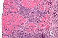
Chloroma - intermed. mag. (WC)
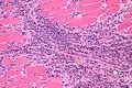
Chloroma - high mag. (WC)
Chloroma - very high mag. (WC)
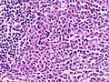
Myeloid sarcoma within a lymph node (WC)


www:
- Granulocytic sarcoma - several crappy images (upmc.edu).
- Myeloid sarcoma - several images (upmc.edu).
IHC
Features:[4] Markers of immaturity:
- CD34 +ve/-ve (5 of 9 cases).
- CD117 +ve (9 of 9 cases).
- TdT
Myeloid markers:
- CD43 +ve (7 of 7 cases) - sensitive, but not specific
- Myeloperoxidase +ve (8 of 10 cases).
- CD11c (myelomonocytic marker)
- CD13 (granulopoietic marker)
- CD33 (granulopoietic marker, specific but less sensitive)
CD34, CD117 and myeloperoxidase are more commonly positive in cases showing granulopoietic differentiation, but can be negative in cases with a myelomonocytic or monocytic differentiation, where CD68, CD163 and lysozyme may be helpful.[5]
Sign out
- It is prudent to mention acute myeloid leukemia somewhere in the report to ensure the appropriate referral is made.
See also
References
- ↑ Bakst, RL.; Tallman, MS.; Douer, D.; Yahalom, J. (Oct 2011). "How I treat extramedullary acute myeloid leukemia.". Blood 118 (14): 3785-93. doi:10.1182/blood-2011-04-347229. PMID 21795742.
- ↑ 2.0 2.1 Eom, KS.; Kim, TY. (Mar 2011). "Intraparenchymal myeloid sarcoma and subsequent spinal myeloid sarcoma for acute myeloblastic leukemia.". J Korean Neurosurg Soc 49 (3): 171-4. doi:10.3340/jkns.2011.49.3.171. PMC 3085814. PMID 21556238. http://www.ncbi.nlm.nih.gov/pmc/articles/PMC3085814/.
- ↑ Arthur, C.; Cermak, J.; Delaunay, J.; Mayer, J.; Mazur, G.; Thomas, X.; Wierzbowska, A.; Jones, MM. et al. (2015). "Post hoc analysis of the relationship between baseline white blood cell count and survival outcome in a randomized Phase III trial of decitabine in older patients with newly diagnosed acute myeloid leukemia.". J Blood Med 6: 25-9. doi:10.2147/JBM.S64067. PMID 25678833.
- ↑ Seifert, RP.; Bulkeley, W.; Zhang, L.; Menes, M.; Bui, MM. (Aug 2014). "A practical approach to diagnose soft tissue myeloid sarcoma preceding or coinciding with acute myeloid leukemia.". Ann Diagn Pathol 18 (4): 253-60. doi:10.1016/j.anndiagpath.2014.06.001. PMID 24969631.
- ↑ "Myeloid sarcoma of the head and neck region". Arch Pathol Lab Med 137 (11): 1560–8. November 2013. doi:10.5858/arpa.2012-0537-OA. PMID 23530613.