Difference between revisions of "Granular cell tumour"
Jump to navigation
Jump to search
| Line 99: | Line 99: | ||
Images: | Images: | ||
*[http://pathhsw5m54.ucsf.edu/cts/grancell.html Granular cell tumour - several LM and EM images (ucsf.edu)]. | *[http://pathhsw5m54.ucsf.edu/cts/grancell.html Granular cell tumour - several LM and EM images (ucsf.edu)]. | ||
==Sign out== | |||
<pre> | |||
SKIN LESION, NECK, EXCISION: | |||
- GRANULAR CELL TUMOUR. | |||
- MILD SOLAR ELASTOSIS. | |||
COMMENT: | |||
The lesional cells stain as follows: | |||
POSITIVE: S-100, CD68, vimentin, calretinin. | |||
NEGATIVE: HMB-45. | |||
</pre> | |||
===Micro=== | |||
The sections show skin with nests of cells in the mid dermis, extending to the subcutis. The cells that make up the nests have an abundant granular grey cytoplasm, and round regular nuclei without obvious nucleoli. No mitotic activity is readily apparent. The lesional cells focally wrap around nerve. The superficial dermis has mild solar elastosis. The lesion is completely excised in the plane of section. | |||
==See also== | ==See also== | ||
Revision as of 12:32, 24 June 2014
| Granular cell tumour | |
|---|---|
| Diagnosis in short | |
_skin.jpg) Granular cell tumour. H&E stain. | |
|
| |
| LM | cells with abundant eosinophilic granular cytoplasm (granules ~ 1-3 micrometers, poorly demarcated on LM), +/-pseudoepitheliomatous hyperplasia |
| LM DDx | squamous cell carcinoma, oncocytoma, adjacent ulcer, xanthoma, melanocytic nevus with neurotization |
| Stains | PAS +ve |
| IHC | S-100 +ve, CD68 +ve (cytoplasmic), vimentin +ve (membranous), calretinin +ve (usually) |
| EM | abundant lysosomes |
| Gross | yellow nodule |
| Site | typically head and neck - other sites: breast, skin, tongue, esophagus + more |
|
| |
| Syndromes | LEOPARD syndrome |
|
| |
| Prevalence | rare |
| Prognosis | usu. benign, may be malignant |
The granular cell tumour is a rare histomorphologically distinctive neoplasm found at many sites. The classic location is the head and neck.
General
- Rare.
- Usually benign.
- May seen in the context of LEOPARD syndrome and a mutation in the PTPN11 gene.[1]
- PTPN11 = protein-tyrosine phosphatase non-receptor type 11.[2]
- Gene implicated in Noonan syndrome 1.
- PTPN11 = protein-tyrosine phosphatase non-receptor type 11.[2]
- May mimic (well-differentiated) squamous cell carcinoma - histopathologically.
- There is a well-described phenomenon called pseudoepitheliomatous hyperplasia.[3]
Aside:
- Pseudoepitheliomatous hyperplasia is seen in:
- Fungal infections.
- Inflammatory papillary hyperplasia.
- Granular cell tumour.
- Adjacent to an ulcer.
Sites
May be seen in any number of sites:
- Granular cell tumour of the breast.
- Granular cell tumour of the skin.
- Granular cell tumour of the tongue.
- Granular cell tumour of the esophagus.
Gross
- Yellow nodule.
DDx of yellow nodule:
- Granular cell tumour.
- Lipoma.
- Xanthoma.
Microscopic
Features:
- Cells with abundant eosinophilic granular cytoplasm - key feature.
- Granules (represent abundant lysosomes[4]):
- Size: 1-3 micrometers.
- Poorly demarcated (on light microscopy).
- Granules (represent abundant lysosomes[4]):
- Nested architecture.
- +/-Pseudoepitheliomatous hyperplasia.
- May mimic SCC.
DDx:
- Squamous cell carcinoma.
- Oncocytoma.
- Melanocytic nevus with neurotization.
- Xanthoma.
Special stains
- PAS +ve.
IHC
Features:[5]
- S100 +ve.
- CD68 +ve (cytoplasmic).
- Vimentin +ve (membranous).
- Calretinin +ve (90-95%).[6]
Images
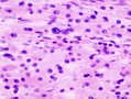
GCT. (WC)
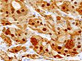
GCT - S-100. (WC)
_S-100.JPG)
EM
- Abundant lysosomes.[4]
- Round structures with variable (electron) density.
Images:
Sign out
SKIN LESION, NECK, EXCISION:
- GRANULAR CELL TUMOUR.
- MILD SOLAR ELASTOSIS.

COMMENT:
The lesional cells stain as follows:
POSITIVE: S-100, CD68, vimentin, calretinin.
NEGATIVE: HMB-45.
Micro
The sections show skin with nests of cells in the mid dermis, extending to the subcutis. The cells that make up the nests have an abundant granular grey cytoplasm, and round regular nuclei without obvious nucleoli. No mitotic activity is readily apparent. The lesional cells focally wrap around nerve. The superficial dermis has mild solar elastosis. The lesion is completely excised in the plane of section.
See also
References
- ↑ Schrader, KA.; Nelson, TN.; De Luca, A.; Huntsman, DG.; McGillivray, BC. (Feb 2009). "Multiple granular cell tumors are an associated feature of LEOPARD syndrome caused by mutation in PTPN11.". Clin Genet 75 (2): 185-9. doi:10.1111/j.1399-0004.2008.01100.x. PMID 19054014.
- ↑ Online 'Mendelian Inheritance in Man' (OMIM) 176876
- ↑ Abu-Eid R, Landini G (March 2006). "Morphometrical differences between pseudoepitheliomatous hyperplasia in granular cell tumours and squamous cell carcinomas". Histopathology 48 (4): 407–16. doi:10.1111/j.1365-2559.2006.02350.x. PMID 16487362.
- ↑ 4.0 4.1 Ordóñez, NG. (Jul 1999). "Granular cell tumor: a review and update.". Adv Anat Pathol 6 (4): 186-203. PMID 10410172.
- ↑ Rekhi, B.; Jambhekar, NA. (Jun 2010). "Morphologic spectrum, immunohistochemical analysis, and clinical features of a series of granular cell tumors of soft tissues: a study from a tertiary referral cancer center.". Ann Diagn Pathol 14 (3): 162-7. doi:10.1016/j.anndiagpath.2010.01.005. PMID 20471560.
- ↑ Fine, SW.; Li, M. (Feb 2003). "Expression of calretinin and the alpha-subunit of inhibin in granular cell tumors.". Am J Clin Pathol 119 (2): 259-64. doi:10.1309/GRH4-JWX6-J9J7-QQTA. PMID 12579997.