Difference between revisions of "Granular cell tumour"
Jump to navigation
Jump to search
(→Images) |
(+infobox) |
||
| Line 1: | Line 1: | ||
{{ Infobox diagnosis | |||
| Name = {{PAGENAME}} | |||
| Image = Granular_cell_tumor_(3)_skin.jpg | |||
| Width = | |||
| Caption = Granular cell tumour. [[H&E stain]]. | |||
| Micro = cells with abundant eosinophilic granular cytoplasm (granules ~ 1-3 micrometers, poorly demarcated on LM), +/-[[pseudoepitheliomatous hyperplasia]] | |||
| Subtypes = | |||
| LMDDx = [[squamous cell carcinoma]], [[oncocytoma]], adjacent ulcer | |||
| Stains = PAS +ve | |||
| IHC = S-100 +ve, CD68 +ve (cytoplasmic), vimentin +ve (membranous), calretinin +ve (usually) | |||
| EM = abundant lysosomes | |||
| Molecular = | |||
| IF = | |||
| Gross = yellow nodule | |||
| Grossing = | |||
| Site = typically [[head and neck pathology|head and neck]] - other sites: [[breast]], [[skin]], tongue, [[esophagus]] + more | |||
| Assdx = | |||
| Syndromes = [[LEOPARD syndrome]] | |||
| Clinicalhx = | |||
| Signs = | |||
| Symptoms = | |||
| Prevalence = rare | |||
| Bloodwork = | |||
| Rads = | |||
| Endoscopy = | |||
| Prognosis = usu. benign, may be malignant | |||
| Other = | |||
| ClinDDx = | |||
}} | |||
The '''granular cell tumour''' is a rare histomorphologically distinctive neoplasm found at many sites. The classic location is the head and neck. | The '''granular cell tumour''' is a rare histomorphologically distinctive neoplasm found at many sites. The classic location is the head and neck. | ||
Revision as of 15:05, 28 September 2013
| Granular cell tumour | |
|---|---|
| Diagnosis in short | |
_skin.jpg) Granular cell tumour. H&E stain. | |
|
| |
| LM | cells with abundant eosinophilic granular cytoplasm (granules ~ 1-3 micrometers, poorly demarcated on LM), +/-pseudoepitheliomatous hyperplasia |
| LM DDx | squamous cell carcinoma, oncocytoma, adjacent ulcer |
| Stains | PAS +ve |
| IHC | S-100 +ve, CD68 +ve (cytoplasmic), vimentin +ve (membranous), calretinin +ve (usually) |
| EM | abundant lysosomes |
| Gross | yellow nodule |
| Site | typically head and neck - other sites: breast, skin, tongue, esophagus + more |
|
| |
| Syndromes | LEOPARD syndrome |
|
| |
| Prevalence | rare |
| Prognosis | usu. benign, may be malignant |
The granular cell tumour is a rare histomorphologically distinctive neoplasm found at many sites. The classic location is the head and neck.
General
- Rare.
- Usually benign.
- May seen in the context of LEOPARD syndrome and a mutation in the PTPN11 gene.[1]
- PTPN11 = protein-tyrosine phosphatase non-receptor type 11.[2]
- Gene implicated in Noonan syndrome 1.
- PTPN11 = protein-tyrosine phosphatase non-receptor type 11.[2]
- May mimic (well-differentiated) squamous cell carcinoma - histopathologically.
- There is a well-described phenomenon called pseudoepitheliomatous hyperplasia.[3]
Aside:
- Pseudoepitheliomatous hyperplasia is seen in:
- Fungal infections.
- Inflammatory papillary hyperplasia.
- Granular cell tumour.
- Adjacent to an ulcer.
Sites
May be seen in any number of sites:
- Granular cell tumour of the breast.
- Granular cell tumour of the skin.
- Granular cell tumour of the tongue.
- Granular cell tumour of the esophagus.
Gross
- Yellow nodule.
DDx of yellow nodule:
- Granular cell tumour.
- Lipoma.
- Xanthoma.
Microscopic
Features:
- Cells with abundant eosinophilic granular cytoplasm - key feature.
- Granules (represent abundant lysosomes[4]):
- Size: 1-3 micrometers.
- Poorly demarcated (on light microscopy).
- Granules (represent abundant lysosomes[4]):
- Nested architecture.
- +/-Pseudoepitheliomatous hyperplasia.
- May mimic SCC.
DDx:
Special stains
- PAS +ve.
IHC
Features:[5]
- S100 +ve.
- CD68 +ve (cytoplasmic).
- Vimentin +ve (membranous).
- Calretinin +ve (90-95%).[6]
Images
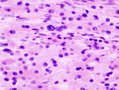
GCT. (WC)
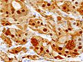
GCT - S-100. (WC)
_S-100.JPG)
EM
- Abundant lysosomes.[4]
- Round structures with variable (electron) density.
Images:
See also
References
- ↑ Schrader, KA.; Nelson, TN.; De Luca, A.; Huntsman, DG.; McGillivray, BC. (Feb 2009). "Multiple granular cell tumors are an associated feature of LEOPARD syndrome caused by mutation in PTPN11.". Clin Genet 75 (2): 185-9. doi:10.1111/j.1399-0004.2008.01100.x. PMID 19054014.
- ↑ Online 'Mendelian Inheritance in Man' (OMIM) 176876
- ↑ Abu-Eid R, Landini G (March 2006). "Morphometrical differences between pseudoepitheliomatous hyperplasia in granular cell tumours and squamous cell carcinomas". Histopathology 48 (4): 407–16. doi:10.1111/j.1365-2559.2006.02350.x. PMID 16487362.
- ↑ 4.0 4.1 Ordóñez, NG. (Jul 1999). "Granular cell tumor: a review and update.". Adv Anat Pathol 6 (4): 186-203. PMID 10410172.
- ↑ Rekhi, B.; Jambhekar, NA. (Jun 2010). "Morphologic spectrum, immunohistochemical analysis, and clinical features of a series of granular cell tumors of soft tissues: a study from a tertiary referral cancer center.". Ann Diagn Pathol 14 (3): 162-7. doi:10.1016/j.anndiagpath.2010.01.005. PMID 20471560.
- ↑ Fine, SW.; Li, M. (Feb 2003). "Expression of calretinin and the alpha-subunit of inhibin in granular cell tumors.". Am J Clin Pathol 119 (2): 259-64. doi:10.1309/GRH4-JWX6-J9J7-QQTA. PMID 12579997.