Difference between revisions of "Giardiasis"
Jump to navigation
Jump to search
(redirect, +cat.) |
(→www: mk nicer) |
||
| (10 intermediate revisions by the same user not shown) | |||
| Line 1: | Line 1: | ||
{{ Infobox diagnosis | |||
| Name = {{PAGENAME}} | |||
| Image = Giardia_small_bowel_--_high_mag.jpg | |||
| Width = | |||
| Caption = Giardiasis. [[H&E stain]]. | |||
| Synonyms = | |||
| Micro = flagellate protozoa - pale/transluence on H&E, 12-15 micrometers (long axis) x 6-10 micrometers (short axis); +/-loss of villi, intraepithelial lymphocytes, inflammatory cells - especially close to the luminal surface | |||
| Subtypes = | |||
| LMDDx = [[celiac disease]] | |||
| Stains = methylene blue +ve | |||
| IHC = CD117 +ve (microorganisms) | |||
| EM = | |||
| Molecular = | |||
| IF = | |||
| Gross = | |||
| Grossing = | |||
| Site = [[duodenum]] | |||
| Assdx = | |||
| Syndromes = | |||
| Clinicalhx = | |||
| Signs = diarrhea x5 days, foul smelling feces, flatulence | |||
| Symptoms = fatigue, abdominal cramps, nausea | |||
| Prevalence = uncommon | |||
| Bloodwork = | |||
| Rads = | |||
| Endoscopy = +/-white spots, diffuse changes | |||
| Prognosis = good, benign | |||
| Other = | |||
| ClinDDx = celiac disease, other causes of diarrhea | |||
| Tx = antibiotics | |||
}} | |||
'''Giardiasis''' is a rare infection, classically found in the [[duodenum]]. It can mimic [[celiac disease]]. It is also known as '''beaver fever'''. | |||
==General== | |||
Clinical features - usually two or more of the following:<ref name=pmid1994703>{{Cite journal | last1 = Hopkins | first1 = RS. | last2 = Juranek | first2 = DD. | title = Acute giardiasis: an improved clinical case definition for epidemiologic studies. | journal = Am J Epidemiol | volume = 133 | issue = 4 | pages = 402-7 | month = Feb | year = 1991 | doi = | PMID = 1994703 }}</ref> | |||
*Diarrhea - x5 days. | |||
*Flatulence. | |||
*Foul smelling feces. | |||
*Nausea. | |||
*Abdominal cramps. | |||
*Excessive tiredness. | |||
Epidemiology: | |||
*Uncommon. | |||
Etiology: | |||
*Flagellate protozoan ''Giardia lamblia''. | |||
Treatment | |||
*Antibiotics, e.g. metronidazole (Flagyl). | |||
==Gross== | |||
*Diffuse changes. | |||
*May have scattered white spots.<ref name=pmid19906109>{{Cite journal | last1 = Biyikoğlu | first1 = I. | last2 = Babali | first2 = A. | last3 = Cakal | first3 = B. | last4 = Köklü | first4 = S. | last5 = Filik | first5 = L. | last6 = Astarci | first6 = MH. | last7 = Ustün | first7 = H. | last8 = Ustündağ | first8 = Y. | last9 = Akbal | first9 = E. | title = Do scattered white spots in the duodenum mark a specific gastrointestinal pathology? | journal = J Dig Dis | volume = 10 | issue = 4 | pages = 300-4 | month = Nov | year = 2009 | doi = 10.1111/j.1751-2980.2009.00399.x | PMID = 19906109 | URL = http://onlinelibrary.wiley.com/doi/10.1111/j.1751-2980.2009.00399.x/pdf }}</ref> | |||
==Microscopic== | |||
Features: | |||
*+/-Loss of villi. | |||
*[[Intraepithelial lymphocytes]]. | |||
**+Other inflammatory cells, especially [[PMNs]], close to the luminal surface. | |||
*Flagellate protozoa -- '''diagnostic feature'''. | |||
**Organisms often at site of bad inflammation. | |||
**Pale/translucent on H&E. | |||
**Size: 12-15 micrometers (long axis) x 6-10 micrometers (short axis) -- if seen completely.<ref>[http://www.water-research.net/Giardia.htm http://www.water-research.net/Giardia.htm]</ref> | |||
***Often look like a crescent moon ([http://en.wikipedia.org/wiki/File:Crescent_Moon.JPG image of crescent moon]) or semicircular<ref>[http://en.wikipedia.org/wiki/Semicircle http://en.wikipedia.org/wiki/Semicircle]</ref> -- as the long axis of the organism is rarely in the plane of the (histologic) section. | |||
Note: | |||
*Changes are typically diffuse, i.e. if multiple biopsies are done the changes are present in all fragments.<ref name=pmid18354756>{{Cite journal | last1 = Freeman | first1 = HJ. | title = Pearls and pitfalls in the diagnosis of adult celiac disease. | journal = Can J Gastroenterol | volume = 22 | issue = 3 | pages = 273-80 | month = Mar | year = 2008 | doi = | PMID = 18354756 }}</ref> | |||
DDx: | |||
*[[Celiac disease]] - near perfect mimic; missing giardia organisms. | |||
===Images=== | |||
====Case 1==== | |||
<gallery> | |||
Image:Giardiasis_duodenum_high.jpg | Giardiasis - high mag. (WC) | |||
Image:Giardiasis_duodenum_low.jpg | Giardiasis - low mag. (WC) | |||
</gallery> | |||
====Case 2==== | |||
<gallery> | |||
Giardia small bowel -- low mag.jpg | Giardia - low mag. | |||
Giardia small bowel -- intermed mag.jpg | Giardia - intermed. mag. | |||
Giardia small bowel -- high mag.jpg | Giardia - high mag. | |||
Giardia small bowel - alt -- high mag.jpg | Giardia - high mag. | |||
Giardia small bowel -- very high mag.jpg | Giardia - very high mag. | |||
</gallery> | |||
====www==== | |||
*[http://path.upmc.edu/cases/case278.html Giardiasis - several images (upmc.edu)]. | |||
==Stains== | |||
*Methylene blue +ve.<ref name=pmid23285438>{{Cite journal | last1 = Rajurkar | first1 = MN. | last2 = Lall | first2 = N. | last3 = Basak | first3 = S. | last4 = Mallick | first4 = SK. | title = A simple method for demonstrating the giardia lamblia trophozoite. | journal = J Clin Diagn Res | volume = 6 | issue = 9 | pages = 1492-4 | month = Nov | year = 2012 | doi = 10.7860/JCDR/2012/4358.2541 | PMID = 23285438 }}</ref> | |||
==IHC== | |||
*CD117 +ve.<ref name=pmid18835628>{{Cite journal | last1 = Sinelnikov | first1 = I. | last2 = Sion-Vardy | first2 = N. | last3 = Shaco-Levy | first3 = R. | title = C-kit (CD117) immunostain is useful for the diagnosis of Giardia lamblia in duodenal biopsies. | journal = Hum Pathol | volume = 40 | issue = 3 | pages = 323-5 | month = Mar | year = 2009 | doi = 10.1016/j.humpath.2008.07.015 | PMID = 18835628 }}</ref> | |||
==Sign out== | |||
<pre> | |||
A. Duodenum, Biopsy: | |||
- Abundant micro-organisms consistent with GIARDIA and small | |||
bowel mucosa with increased intraepithelial lymphocytes, see comment. | |||
- NEGATIVE for dysplasia. | |||
B. Stomach, Biopsy: | |||
- Body and antral-type mucosa with mild chronic inactive inflammation. | |||
- NEGATIVE for Helicobacter-like organisms. | |||
- NEGATIVE for intestinal metaplasia. | |||
- NEGATIVE for dysplasia and NEGATIVE for malignancy. | |||
Comment: | |||
The increased intraepithelial lymphocytes are likely to due to the Giardia; however, other | |||
causes cannot be excluded. | |||
</pre> | |||
===Block letters=== | |||
<pre> | |||
DUODENUM, BIOPSY: | |||
- SMALL BOWEL MUCOSA WITH BRUNNER'S GLANDS AND MICROORGANISMS CONSISTENT WITH GIARDIA. | |||
</pre> | |||
==See also== | |||
*[[Duodenum]]. | |||
==References== | |||
{{Reflist|2}} | |||
[[Category:Diagnosis]] | [[Category:Diagnosis]] | ||
[[Category:Duodenum]] | |||
Latest revision as of 21:32, 14 July 2019
| Giardiasis | |
|---|---|
| Diagnosis in short | |
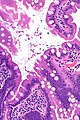
 Giardiasis. H&E stain. | |
|
| |
| LM | flagellate protozoa - pale/transluence on H&E, 12-15 micrometers (long axis) x 6-10 micrometers (short axis); +/-loss of villi, intraepithelial lymphocytes, inflammatory cells - especially close to the luminal surface |
| LM DDx | celiac disease |
| Stains | methylene blue +ve |
| IHC | CD117 +ve (microorganisms) |
| Site | duodenum |
|
| |
| Signs | diarrhea x5 days, foul smelling feces, flatulence |
| Symptoms | fatigue, abdominal cramps, nausea |
| Prevalence | uncommon |
| Endoscopy | +/-white spots, diffuse changes |
| Prognosis | good, benign |
| Clin. DDx | celiac disease, other causes of diarrhea |
| Treatment | antibiotics |
Giardiasis is a rare infection, classically found in the duodenum. It can mimic celiac disease. It is also known as beaver fever.
General
Clinical features - usually two or more of the following:[1]
- Diarrhea - x5 days.
- Flatulence.
- Foul smelling feces.
- Nausea.
- Abdominal cramps.
- Excessive tiredness.
Epidemiology:
- Uncommon.
Etiology:
- Flagellate protozoan Giardia lamblia.
Treatment
- Antibiotics, e.g. metronidazole (Flagyl).
Gross
- Diffuse changes.
- May have scattered white spots.[2]
Microscopic
Features:
- +/-Loss of villi.
- Intraepithelial lymphocytes.
- +Other inflammatory cells, especially PMNs, close to the luminal surface.
- Flagellate protozoa -- diagnostic feature.
- Organisms often at site of bad inflammation.
- Pale/translucent on H&E.
- Size: 12-15 micrometers (long axis) x 6-10 micrometers (short axis) -- if seen completely.[3]
- Often look like a crescent moon (image of crescent moon) or semicircular[4] -- as the long axis of the organism is rarely in the plane of the (histologic) section.
Note:
- Changes are typically diffuse, i.e. if multiple biopsies are done the changes are present in all fragments.[5]
DDx:
- Celiac disease - near perfect mimic; missing giardia organisms.
Images
Case 1
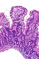
Giardiasis - high mag. (WC)
Giardiasis - low mag. (WC)


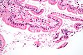
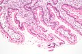
Case 2
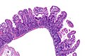
Giardia - low mag.
Giardia - intermed. mag.
Giardia - high mag.

Giardia - high mag.
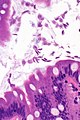
Giardia - very high mag.


www
Stains
- Methylene blue +ve.[6]
IHC
- CD117 +ve.[7]
Sign out
A. Duodenum, Biopsy:
- Abundant micro-organisms consistent with GIARDIA and small
  bowel mucosa with increased intraepithelial lymphocytes, see comment.
- NEGATIVE for dysplasia.

B. Stomach, Biopsy:
- Body and antral-type mucosa with mild chronic inactive inflammation.
- NEGATIVE for Helicobacter-like organisms.
- NEGATIVE for intestinal metaplasia.
- NEGATIVE for dysplasia and NEGATIVE for malignancy.

Comment:
The increased intraepithelial lymphocytes are likely to due to the Giardia; however, other
causes cannot be excluded.
Block letters
DUODENUM, BIOPSY:
- SMALL BOWEL MUCOSA WITH BRUNNER'S GLANDS AND MICROORGANISMS CONSISTENT WITH GIARDIA.
See also
References
- ↑ Hopkins, RS.; Juranek, DD. (Feb 1991). "Acute giardiasis: an improved clinical case definition for epidemiologic studies.". Am J Epidemiol 133 (4): 402-7. PMID 1994703.
- ↑ Biyikoğlu, I.; Babali, A.; Cakal, B.; Köklü, S.; Filik, L.; Astarci, MH.; Ustün, H.; Ustündağ, Y. et al. (Nov 2009). "Do scattered white spots in the duodenum mark a specific gastrointestinal pathology?". J Dig Dis 10 (4): 300-4. doi:10.1111/j.1751-2980.2009.00399.x. PMID 19906109.
- ↑ http://www.water-research.net/Giardia.htm
- ↑ http://en.wikipedia.org/wiki/Semicircle
- ↑ Freeman, HJ. (Mar 2008). "Pearls and pitfalls in the diagnosis of adult celiac disease.". Can J Gastroenterol 22 (3): 273-80. PMID 18354756.
- ↑ Rajurkar, MN.; Lall, N.; Basak, S.; Mallick, SK. (Nov 2012). "A simple method for demonstrating the giardia lamblia trophozoite.". J Clin Diagn Res 6 (9): 1492-4. doi:10.7860/JCDR/2012/4358.2541. PMID 23285438.
- ↑ Sinelnikov, I.; Sion-Vardy, N.; Shaco-Levy, R. (Mar 2009). "C-kit (CD117) immunostain is useful for the diagnosis of Giardia lamblia in duodenal biopsies.". Hum Pathol 40 (3): 323-5. doi:10.1016/j.humpath.2008.07.015. PMID 18835628.