Difference between revisions of "Giant cells"
Jump to navigation
Jump to search
(→Table) |
(→Table) |
||
| (4 intermediate revisions by the same user not shown) | |||
| Line 19: | Line 19: | ||
|- | |- | ||
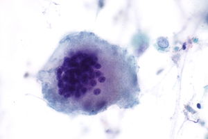
| Touton giant cell | | Touton giant cell | ||
| | | Nuclei form a ring around the cell periphery with eosinophilic cytoplasm centrally and foamy cytoplasm at the periphery. | ||
| [[ | | [[Juvenile xanthogranuloma]], [[xanthoma]], [[Erdheim-Chester disease]], [[fat necrosis]], [[dermatofibroma]] | ||
| | | High lipid content lesions<ref>URL: [http://granuloma.homestead.com/giant_cells.html http://granuloma.homestead.com/giant_cells.html]. Accessed on: 7 February 2011.</ref>, Named after Karl Touton | ||
| [[Image:Juvenile_xanthogranuloma_-_very_high_mag.jpg|thumb|200px|JXG (WC)]] | | [[Image:Juvenile_xanthogranuloma_-_very_high_mag.jpg|thumb|200px|JXG (WC)]] | ||
|- | |- | ||
| Line 31: | Line 31: | ||
|- | |- | ||
| Langhans giant cell | | Langhans giant cell | ||
| peripheral eccentric nuclei<ref name=Ref_InstantPath7>{{Ref InstantPath|7}}</ref> | | peripheral semi-circular eccentric nuclei<ref name=Ref_InstantPath7>{{Ref InstantPath|7}}</ref> | ||
| | | tuberculosis, sarcoidosis. | ||
| '''not''' to be confused with ''Langerhans cells'' | | '''not''' to be confused with ''Langerhans cells'', Named after Theodor Langhans | ||
| [[Image:Granulation_tissue_containg_a_poorly_formed_granuloma_with_a_Langhan%27s_giant_cell.jpg|thumb|200px|LGC (WC)]] | | [[Image:Granulation_tissue_containg_a_poorly_formed_granuloma_with_a_Langhan%27s_giant_cell.jpg|thumb|200px|LGC (WC)]] | ||
|- | |- | ||
| Osteoclast-like giant cells | | Osteoclast-like giant cells | ||
| | | multiple bland central nuclei, ruffled cell membrane. | ||
| osteoclasts, others | | osteoclasts, others | ||
| [[AKA]] osteoclast-type giant cells | | [[AKA]] osteoclast-type giant cells | ||
Latest revision as of 09:18, 13 August 2018
Giant cells are "big" cells with multiple nuclei. They come in different flavours, which are suggestive of causality.

This article deals with the classic types of giant cells. A more general differential diagnosis of giant cells is in giant cell lesions.
Giant cell types
List:
- Touton giant cell.
- Osteoclast-like giant cell.
- Foreign body type giant cell.
Table
| Type | Histology | DDx | Other | Image |
| Touton giant cell | Nuclei form a ring around the cell periphery with eosinophilic cytoplasm centrally and foamy cytoplasm at the periphery. | Juvenile xanthogranuloma, xanthoma, Erdheim-Chester disease, fat necrosis, dermatofibroma | High lipid content lesions[1], Named after Karl Touton | |
| Epithelioid type | scattered nuclei[2] | drug reaction, neoplasm, foreign body, infection, idiopathic, autoimmune, allergic | granulomatous inflammation | |
| Langhans giant cell | peripheral semi-circular eccentric nuclei[2] | tuberculosis, sarcoidosis. | not to be confused with Langerhans cells, Named after Theodor Langhans | |
| Osteoclast-like giant cells | multiple bland central nuclei, ruffled cell membrane. | osteoclasts, others | AKA osteoclast-type giant cells |


See also
- Basics.
- Giant cell lesions - includes a DDx of lesions with giant cells.
- Histiocytoses.
References
- ↑ URL: http://granuloma.homestead.com/giant_cells.html. Accessed on: 7 February 2011.
- ↑ 2.0 2.1 Borley, Neil R.; Warren, Bryan F. (2007). Instant Pathology (1st ed.). Wiley-Blackwell. pp. 7. ISBN 978-1405132909.