Difference between revisions of "Giant cell cystitis"
Jump to navigation
Jump to search
| (9 intermediate revisions by the same user not shown) | |||
| Line 1: | Line 1: | ||
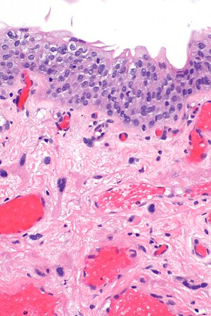
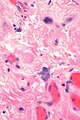
'''Giant cell cystitis''' is term used for a benign change of | [[Image:Bladder with benign large mesenchymal cells -- high mag.jpg|thumb|right|300px|Urinary bladder with benign large mesenchymal cells - so-called "giant cell cystitis". [[H&E stain]].]] | ||
'''Giant cell cystitis''' is term used for a benign change of mesenchymal cells in the [[urinary bladder]] lamina propria. | |||
''Giant cell cystitis'' is considered a misnomer as may be seen in an otherwise normal bladder.<ref name=pmid20196670>{{Cite journal | last1 = Hameed | first1 = O. | last2 = Humphrey | first2 = PA. | title = Pseudoneoplastic mimics of prostate and bladder carcinomas. | journal = Arch Pathol Lab Med | volume = 134 | issue = 3 | pages = 427-43 | month = Mar | year = 2010 | doi = 10.1043/1543-2165-134.3.427 | PMID = 20196670 }}</ref> | ''Giant cell cystitis'' is considered a misnomer as it may be seen in an otherwise normal bladder that lacks significant inflammation.<ref name=pmid20196670>{{Cite journal | last1 = Hameed | first1 = O. | last2 = Humphrey | first2 = PA. | title = Pseudoneoplastic mimics of prostate and bladder carcinomas. | journal = Arch Pathol Lab Med | volume = 134 | issue = 3 | pages = 427-43 | month = Mar | year = 2010 | doi = 10.1043/1543-2165-134.3.427 | PMID = 20196670 }}</ref> | ||
==General== | ==General== | ||
*Considered a common benign finding; ''not'' a clinical entity.<ref name=Ref_Amin2_6>{{Ref Amin|2:6}}</ref> | *Considered a common benign finding; ''not'' a clinical entity.<ref name=Ref_Amin2_6>{{Ref Amin|2:6}}</ref> | ||
*Reported in up to 1/3 of bladders at [[autopsy]].<ref>{{Cite journal | last1 = Wells | first1 = HG | last2 = | first2 = | title = Giant cells in cystitis | journal = Arch Pathol | volume = 26 | issue = | pages = 32-43 | month = | year = 1938 | doi = | PMID = | PMC = | url = }}</ref> | |||
==Microscopic== | ==Microscopic== | ||
Features:<ref name=Ref_Uropath305>{{Ref Uropath|305}}</ref> | Features:<ref name=Ref_Uropath305>{{Ref Uropath|305}}</ref> | ||
*Scattered atypical mesenchymal cells - mononuclear or multinucleated. | *Scattered atypical mesenchymal cells - mononuclear or multinucleated - '''key feature'''. | ||
*+/-Nuclear hyperchromasia and/or lobulation. | *+/-Nuclear hyperchromasia and/or lobulation. | ||
* | *Minimal/absence of mitotic activity. | ||
Note: | |||
*Inflammation may be present or absent; "giant cell cystitis" is a misnomer. | |||
DDx: | DDx: | ||
| Line 16: | Line 21: | ||
*[[Sarcoma]] of the bladder or metastatic sarcoma - typically more cellular.<ref name=Ref_Amin2_6/> | *[[Sarcoma]] of the bladder or metastatic sarcoma - typically more cellular.<ref name=Ref_Amin2_6/> | ||
*Sarcomatoid change in [[urothelial carcinoma]]. | *Sarcomatoid change in [[urothelial carcinoma]]. | ||
===Images=== | |||
<gallery> | |||
Image: Bladder with benign large mesenchymal cells -- very low mag.jpg | GCC - very low mag. (WC) | |||
Image: Bladder with benign large mesenchymal cells -- low mag.jpg | GCC - low mag. (WC) | |||
Image: Bladder with benign large mesenchymal cells -- intermed mag.jpg | GCC - intermed. mag. (WC) | |||
Image: Bladder with benign large mesenchymal cells -- high mag.jpg | GCC - high mag. (WC) | |||
Image: Bladder with benign large mesenchymal cells - alt -- intermed mag.jpg | GCC - intermed. mag. (WC) | |||
Image: Bladder with benign large mesenchymal cells - alt -- high mag.jpg | GCC - high mag. (WC) | |||
Image: Bladder with benign large mesenchymal cells - alt -- very high mag.jpg | GCC - very high mag. (WC) | |||
Image: Bladder with benign large mesenchymal cells -- very high mag.jpg | GCC - very high mag. (WC) | |||
</gallery> | |||
==Sign out== | ==Sign out== | ||
| Line 27: | Line 45: | ||
- NEGATIVE for significant proliferative activity and NEGATIVE for significant inflammation. | - NEGATIVE for significant proliferative activity and NEGATIVE for significant inflammation. | ||
- NEGATIVE for dysplasia and NEGATIVE for malignancy. | - NEGATIVE for dysplasia and NEGATIVE for malignancy. | ||
</pre> | </pre> | ||
==See also== | ==See also== | ||
*[[Radiation cystitis]]. | *[[Radiation cystitis]]. | ||
*[[Giant cells]]. | |||
==References== | ==References== | ||
Latest revision as of 14:12, 7 December 2016

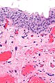
Urinary bladder with benign large mesenchymal cells - so-called "giant cell cystitis". H&E stain.
Giant cell cystitis is term used for a benign change of mesenchymal cells in the urinary bladder lamina propria.
Giant cell cystitis is considered a misnomer as it may be seen in an otherwise normal bladder that lacks significant inflammation.[1]
General
- Considered a common benign finding; not a clinical entity.[2]
- Reported in up to 1/3 of bladders at autopsy.[3]
Microscopic
Features:[4]
- Scattered atypical mesenchymal cells - mononuclear or multinucleated - key feature.
- +/-Nuclear hyperchromasia and/or lobulation.
- Minimal/absence of mitotic activity.
Note:
- Inflammation may be present or absent; "giant cell cystitis" is a misnomer.
DDx:
- Radiation cystitis - history of radiation, more atypical usually with nucleoli.[2]
- Sarcoma of the bladder or metastatic sarcoma - typically more cellular.[2]
- Sarcomatoid change in urothelial carcinoma.
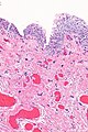
Images
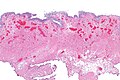
GCC - very low mag. (WC)
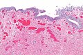
GCC - low mag. (WC)
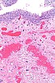
GCC - intermed. mag. (WC)
GCC - high mag. (WC)
GCC - intermed. mag. (WC)
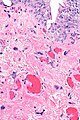
GCC - high mag. (WC)
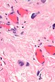
GCC - very high mag. (WC)
GCC - very high mag. (WC)


Sign out
Urinary Bladder, Biopsy:
- Urothelial mucosa with scattered large atypical mesenchymal cells in the
  lamina propria, prominent smooth muscle and prominent superficial
  blood vessels.
- Urothelium within normal limits.
- Thick benign muscularis propria.
- NEGATIVE for significant proliferative activity and NEGATIVE for significant inflammation.
- NEGATIVE for dysplasia and NEGATIVE for malignancy.
See also
References
- ↑ Hameed, O.; Humphrey, PA. (Mar 2010). "Pseudoneoplastic mimics of prostate and bladder carcinomas.". Arch Pathol Lab Med 134 (3): 427-43. doi:10.1043/1543-2165-134.3.427. PMID 20196670.
- ↑ 2.0 2.1 2.2 Amin, Mahul B. (2010). Diagnostic Pathology: Genitourinary (1st ed.). Amirsys. pp. 2:6. ISBN 978-1931884280.
- ↑ Wells, HG (1938). "Giant cells in cystitis". Arch Pathol 26: 32-43.
- ↑ Amin, Mahul B.; Eble, John; Grignon, David; Srigley, John. (2013). Urological Pathology (1st ed.). Wolters Kluwer. pp. 305. ISBN 978-0781782814.