Difference between revisions of "Fuchs dystrophy"
Jump to navigation
Jump to search
(tweak) |
|||
| Line 16: | Line 16: | ||
www: | www: | ||
*[http://www.ncbi.nlm.nih.gov/pmc/articles/PMC2695576/figure/F56/ Fuchs dystrophy (nlm.nih.gov)].<ref name=pmid19236704/> | *[http://www.ncbi.nlm.nih.gov/pmc/articles/PMC2695576/figure/F56/ Fuchs dystrophy (nlm.nih.gov)].<ref name=pmid19236704/> | ||
==Sign out== | |||
<pre> | |||
Decemet's Membrane, Right Eye, Excision: | |||
- Loss of endothelial cells and guttata formation compatible with Fuch's dystrophy. | |||
</pre> | |||
==See also== | ==See also== | ||
Revision as of 16:03, 27 August 2015
Fuchs dystrophy, also Fuchs endothelial dystrophy, is a rare pathology of the eye that may be seen in Descemet's membrane specimens.
General
- Uncommon.
Microscopic
Features:[1]
- Guttae accumulations.
- Collagenous material from the basement membrane.
- Decreased endothelial density.[2]
Images
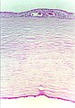
Fuchs dystrophy. (WC)

www:
Sign out
Decemet's Membrane, Right Eye, Excision:
- Loss of endothelial cells and guttata formation compatible with Fuch's dystrophy.
See also
- Eye.
References
- ↑ McLaren, JW.; Bachman, LA.; Kane, KM.; Patel, SV. (Feb 2014). "Objective assessment of the corneal endothelium in Fuchs' endothelial dystrophy.". Invest Ophthalmol Vis Sci 55 (2): 1184-90. doi:10.1167/iovs.13-13041. PMID 24508788.
- ↑ 2.0 2.1 Klintworth, GK. (2009). "Corneal dystrophies.". Orphanet J Rare Dis 4: 7. doi:10.1186/1750-1172-4-7. PMID 19236704.