Difference between revisions of "Fibromatoses"
Jump to navigation
Jump to search
(→Superficial: w) |
|||
| (19 intermediate revisions by the same user not shown) | |||
| Line 1: | Line 1: | ||
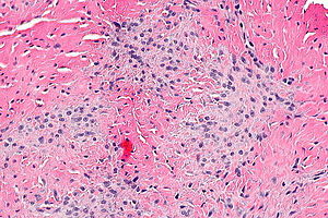
'''Fibromatoses''' are a group of benign stromal lesions that | [[Image:Palmar fibromatosis - alt -- high mag.jpg|thumb|right|Micrograph showing a [[palmar fibromatosis]]. [[H&E stain]].]] | ||
'''Fibromatoses''' are a group of benign stromal lesions that may or may not be associated with [[syndromes]]. | |||
''Plantar fibromatosis'' redirects here. | |||
==Syndromes associated with fibromatoses== | ==Syndromes associated with fibromatoses== | ||
| Line 10: | Line 13: | ||
===Superficial=== | ===Superficial=== | ||
*Palmar ([[Dupuytren's contracture]]). | *Palmar ([[Dupuytren's contracture]]). | ||
*Plantar. | *Plantar (Ledderhose disease<ref>{{cite journal |authors=Mozena JD, Hansen EK, Jones PC |title=Radiotherapy for Plantar Fibromas (Ledderhose Disease) |journal=J Am Podiatr Med Assoc |volume=112 |issue=1 |pages= |date=March 2022 |pmid=35324461 |doi=10.7547/19-008 |url=}}</ref><ref name=pmid35274715>{{cite journal |authors=Schoenfeld JD, Agaram NP, Lefkowitz RA, Kelly CM, Healey JH, Gounder MM |title=Sorafenib in Dupuytren and Ledderhose Disease |journal=Oncologist |volume=27 |issue=3 |pages=e294–e296 |date=March 2022 |pmid=35274715 |pmc=8914480 |doi=10.1093/oncolo/oyab050 |url=}}</ref>). | ||
*[[ | *Penile ([[Peyronie's disease]]). | ||
===Deep=== | ===Deep=== | ||
*[[Desmoid-type fibromatosis]]. | *[[Desmoid-type fibromatosis]]. | ||
*Others. | *Others. | ||
==Gross== | |||
*Firm. | |||
*Infiltrative borders. | |||
Image: | |||
*[http://www.flickr.com/photos/kcrazypathgirl/5858963062/ Desmoid tumour (flickr.com)]. | |||
*[http://www.flickr.com/photos/lunarcaustic/5334047938/ Mesenteric fibromatosis (flickr.com)]. | |||
==Microscopic== | ==Microscopic== | ||
Features: | Features: | ||
*Bland spindle cells - typically in fascicles. | *Bland spindle cells - typically in fascicles. | ||
**Pale eosinophilic cytoplasm.<ref>URL: [http://www.histopathology-india.net/Fibromatosis.htm http://www.histopathology-india.net/Fibromatosis.htm]. Accessed on: 18 September 2012.</ref> | |||
**Nucleus may be ovoid.<ref name=pmid19926768/> | |||
*Small thin blood vessels that are parallel to one another. | *Small thin blood vessels that are parallel to one another. | ||
**Nuclei of [[blood vessel]]s are typically darker staining than those of the lesion. | **Nuclei of [[blood vessel]]s are typically darker staining than those of the lesion. | ||
| Line 26: | Line 39: | ||
*Metaplastic carcinoma, e.g. [[metaplastic breast carcinoma]]. | *Metaplastic carcinoma, e.g. [[metaplastic breast carcinoma]]. | ||
*[[Nodular fasciitis]]. | *[[Nodular fasciitis]]. | ||
*[[Dermatofibroma]]. | |||
*[[GIST]]. | |||
*[[Desmoplastic fibroblastoma]] - esp. shoulder region. | |||
Images: | |||
*[http://www.sarcomaimages.com/index.php?v=Plantar-Fibromatosis Plantar fibromatosis (sarcomaimages.com)]. | |||
*[http://www.flickr.com/photos/lunarcaustic/5333412517/in/photostream/ Mesenteric fibromatosis (flickr.com)]. | |||
*[http://radiographics.rsna.org/content/29/7/2143/F12.expansion.html Plantar fibromatosis (rsna.org)].<ref name=pmid19926768/> | |||
*[http://radiographics.rsna.org/content/29/7/2143/F17.expansion.html Plantar fibromatosis (rsna.org)].<ref name=pmid19926768>{{Cite journal | last1 = Murphey | first1 = MD. | last2 = Ruble | first2 = CM. | last3 = Tyszko | first3 = SM. | last4 = Zbojniewicz | first4 = AM. | last5 = Potter | first5 = BK. | last6 = Miettinen | first6 = M. | title = From the archives of the AFIP: musculoskeletal fibromatoses: radiologic-pathologic correlation. | journal = Radiographics | volume = 29 | issue = 7 | pages = 2143-73 | month = Nov | year = 2009 | doi = 10.1148/rg.297095138 | PMID = 19926768 }}</ref> | |||
==IHC== | |||
*Beta-catenin +ve -- nuclear stain. | |||
*CD117 -ve. | |||
*CD34 -ve.<ref name=pmid25349618>{{Cite journal | last1 = Li Destri | first1 = G. | last2 = Ferraro | first2 = MJ. | last3 = Calabrini | first3 = M. | last4 = Pennisi | first4 = M. | last5 = Magro | first5 = G. | title = Desmoid-type fibromatosis of the mesentery: report of a sporadic case with emphasis on differential diagnostic problems. | journal = Case Rep Med | volume = 2014 | issue = | pages = 850180 | month = | year = 2014 | doi = 10.1155/2014/850180 | PMID = 25349618 }}</ref> | |||
Image: | |||
*[http://pathinfo.wikia.com/wiki/File:Beta-catenin.fibromatosis.1.jpg Fibromatosis (wikia.com)]. | |||
==Sign out== | |||
===Deep=== | |||
<pre> | |||
SOFT TISSUE LESION, LEFT SHOULDER, CORE BIOPSY: | |||
- FIBROMATOSIS, SEE COMMENT. | |||
COMMENT: | |||
The lesion was evaluated with immunostains: | |||
Positive: vimentin, SMA (rare), beta-catenin. | |||
Negative: pankeratin, S-100, desmin, CD34 (stains vessels). | |||
</pre> | |||
===Plantar=== | |||
<pre> | |||
Left Foot, Plantar Fascia, Fasciectomy: | |||
- Plantar fibromatosis. | |||
</pre> | |||
===Micro=== | |||
The sections show a lesion with bland spindle cells. There is no apparent nuclear atypia. Mitotic figures are not identified. No myxoid areas are apparent. No necrosis is identified. | |||
==See also== | ==See also== | ||
| Line 38: | Line 89: | ||
[[Category:Weird stuff]] | [[Category:Weird stuff]] | ||
[[Category:Soft tissue pathology]] | |||
Latest revision as of 17:29, 24 April 2023

Fibromatoses are a group of benign stromal lesions that may or may not be associated with syndromes.
Plantar fibromatosis redirects here.
Syndromes associated with fibromatoses
- Familial adenomatous polyposis.[1]
- Hereditary desmoid syndrome.
Types
Superficial
- Palmar (Dupuytren's contracture).
- Plantar (Ledderhose disease[2][3]).
- Penile (Peyronie's disease).
Deep
- Desmoid-type fibromatosis.
- Others.
Gross
- Firm.
- Infiltrative borders.
Image:
Microscopic
Features:
- Bland spindle cells - typically in fascicles.
- Small thin blood vessels that are parallel to one another.
- Nuclei of blood vessels are typically darker staining than those of the lesion.
DDx:
- Metaplastic carcinoma, e.g. metaplastic breast carcinoma.
- Nodular fasciitis.
- Dermatofibroma.
- GIST.
- Desmoplastic fibroblastoma - esp. shoulder region.
Images:
- Plantar fibromatosis (sarcomaimages.com).
- Mesenteric fibromatosis (flickr.com).
- Plantar fibromatosis (rsna.org).[5]
- Plantar fibromatosis (rsna.org).[5]
IHC
- Beta-catenin +ve -- nuclear stain.
- CD117 -ve.
- CD34 -ve.[6]
Image:
Sign out
Deep
SOFT TISSUE LESION, LEFT SHOULDER, CORE BIOPSY:
- FIBROMATOSIS, SEE COMMENT.

COMMENT:
The lesion was evaluated with immunostains:
Positive: vimentin, SMA (rare), beta-catenin.
Negative: pankeratin, S-100, desmin, CD34 (stains vessels).
Plantar
Left Foot, Plantar Fascia, Fasciectomy:
	- Plantar fibromatosis.
Micro
The sections show a lesion with bland spindle cells. There is no apparent nuclear atypia. Mitotic figures are not identified. No myxoid areas are apparent. No necrosis is identified.
See also
References
- ↑ Kumar, Vinay; Abbas, Abul K.; Fausto, Nelson; Aster, Jon (2009). Robbins and Cotran pathologic basis of disease (8th ed.). Elsevier Saunders. pp. 1092. ISBN 978-1416031215.
- ↑ Mozena JD, Hansen EK, Jones PC (March 2022). "Radiotherapy for Plantar Fibromas (Ledderhose Disease)". J Am Podiatr Med Assoc 112 (1). doi:10.7547/19-008. PMID 35324461.
- ↑ Schoenfeld JD, Agaram NP, Lefkowitz RA, Kelly CM, Healey JH, Gounder MM (March 2022). "Sorafenib in Dupuytren and Ledderhose Disease". Oncologist 27 (3): e294–e296. doi:10.1093/oncolo/oyab050. PMC 8914480. PMID 35274715. https://www.ncbi.nlm.nih.gov/pmc/articles/PMC8914480/.
- ↑ URL: http://www.histopathology-india.net/Fibromatosis.htm. Accessed on: 18 September 2012.
- ↑ 5.0 5.1 5.2 Murphey, MD.; Ruble, CM.; Tyszko, SM.; Zbojniewicz, AM.; Potter, BK.; Miettinen, M. (Nov 2009). "From the archives of the AFIP: musculoskeletal fibromatoses: radiologic-pathologic correlation.". Radiographics 29 (7): 2143-73. doi:10.1148/rg.297095138. PMID 19926768.
- ↑ Li Destri, G.; Ferraro, MJ.; Calabrini, M.; Pennisi, M.; Magro, G. (2014). "Desmoid-type fibromatosis of the mesentery: report of a sporadic case with emphasis on differential diagnostic problems.". Case Rep Med 2014: 850180. doi:10.1155/2014/850180. PMID 25349618.