Difference between revisions of "Fibroepithelial polyp"
(+cat.) |
|||
| (10 intermediate revisions by the same user not shown) | |||
| Line 1: | Line 1: | ||
{{ Infobox diagnosis | |||
| Name = {{PAGENAME}} | |||
| Image = SkinTumors-P9250819.jpg | |||
| Width = | |||
| Caption = Fibroepithelial polyp. [[H&E stain]]. | |||
| Micro = On a stalk / epithelium on three sides, benign epidermis, fibrous core, +/-inflammation | |||
| Subtypes = | |||
| LMDDx = Regressing [[melanocytic lesions]] (esp. [[intradermal melanocytic nevus]]), pedunculated [[seborrheic keratosis]], [[nevus lipomatosus superficialis]], [[neurofibroma]] | |||
| Stains = | |||
| IHC = | |||
| EM = | |||
| Molecular = | |||
| IF = | |||
| Gross = raised skin-coloured lesion | |||
| Grossing = | |||
| Site = [[skin]] | |||
| Assdx = | |||
| Syndromes = [[Birt–Hogg–Dubé syndrome]] | |||
| Clinicalhx = | |||
| Signs = | |||
| Symptoms = | |||
| Prevalence = very common | |||
| Bloodwork = | |||
| Rads = | |||
| Endoscopy = | |||
| Prognosis = benign | |||
| Other = | |||
| ClinDDx = | |||
}} | |||
'''Fibroepithelial polyp''', also known as '''acrochordon''' and '''skin tag''', is a very common benign skin lesion. | |||
The lesion in the mouth is dealt with in the article ''[[oral fibroma]]''. | |||
==General== | |||
*Benign. | |||
*Older people. | |||
*May be associated with pregnancy, [[diabetes]], [[intestinal polyposis]].<ref name=Ref_PCPBoD8|596>{{Ref PCPBoD8|596}}</ref> | |||
*Can be a component of ''[[Birt–Hogg–Dubé syndrome]]''. | |||
May be divided into: | |||
*Epithelial type - abundant epithelium. | |||
*Stromal type - abundant fibrous stroma. | |||
==Gross== | |||
*Raised skin-coloured lesion. | |||
DDx - clinical: | |||
*Skin tag-like nevus. | |||
===Image=== | |||
<gallery> | |||
Image:Skintagblemish.jpg | Skin tag. (WC) | |||
</gallery> | |||
==Microscopic== | |||
Features: | |||
*On a stalk / epithelium on three sides. | |||
*Benign epidermis. | |||
*Fibrous core. | |||
*+/-Inflammation. | |||
DDx: | |||
*Pedunculated [[seborrheic keratosis]] - typically have epidermal hyperplasia, hyperkeratosis, horn cysts.<ref name=Ref_Derm342>{{Ref Derm|342}}</ref> | |||
*Regressing [[melanocytic lesions]]. | |||
**[[Intradermal melanocytic nevus]]. | |||
*[[Nevus lipomatosus superficialis]] - abundant adipocytes in the superficial dermis. | |||
*[[Neurofibroma]]. | |||
*[[Seborrheic keratosis]] - usu. horn cysts. | |||
===Images=== | |||
<gallery> | |||
Image:SkinTumors-P9250819.jpg | Skin tag. (WC) | |||
</gallery> | |||
www: | |||
*[http://dermatlas.med.jhmi.edu/derm/IndexDisplay.cfm?ImageID=1767547949 Fibroepithelial polyp (dermatlas.med.jhmi.edu)].<ref>URL: [http://dermatlas.med.jhmi.edu/derm/result.cfm?Diagnosis=1196583692 http://dermatlas.med.jhmi.edu/derm/result.cfm?Diagnosis=1196583692]. Accessed on: 1 September 2011.</ref> | |||
*[http://www.surgicalpathologyatlas.com/glfusion/mediagallery/media.php?f=0&sort=0&s=20080802171929218 Fibroepithelial polyp (surgicalpathologyatlas.com)]. | |||
*[http://dermatology-s10.cdlib.org/143/case_presentations/skintags/3.jpg Inflamed fibroepithelial polyp (cdlib.org)].<ref>URL: [http://dermatology-s10.cdlib.org/143/case_presentations/skintags/allegue.html http://dermatology-s10.cdlib.org/143/case_presentations/skintags/allegue.html]. Accessed on: 9 January 2013.</ref> | |||
==Sign out== | |||
<pre> | |||
EYELID ("TAG"), RIGHT, REMOVAL: | |||
- BENIGN FIBROEPITHELIAL POLYP. | |||
</pre> | |||
<pre> | |||
SKIN ("SKIN TAG"), THIGH, REMOVAL: | |||
- FIBROEPITHELIAL POLYP. | |||
</pre> | |||
===Micro=== | |||
The sections show a fragment of skin with epithelium on three sides. The epithelium matures normally and is not hypertrophic. Orthokeratosis is present. The core of the lesion has fibrous tissue. There is no significant inflammation. No melanocytic nests are identified. | |||
====Hyperkeratosis==== | |||
The sections show a fragment of skin with epithelium on three sides. The epidermis matures | |||
toward the surface and no significant basilar atypia is identified. A prominent granular layer is present. Mild hyperkeratosis is present. The core of the lesion consists of fibrous tissue. There is no significant inflammation. No | |||
melanocytic nests are identified. | |||
====Inflamed==== | |||
The sections show a fragment of skin with epithelium on three sides. The epithelium matures | |||
and is acanthotic. Minimal parakeratosis is present. The core of the lesion consists of | |||
fibrous tissue with a mild lymphocyte-predominant dermal infiltrate. Rare siderophages are | |||
present. | |||
There is mild basal nuclear enlargement. No significant nuclear atypia is apparent. The | |||
dermal-epidermal interface is well-demarcated. Rare basal mitotic activity is identified. | |||
====Epithelial-type==== | |||
The sections show skin with epithelium on three sides, acanthosis and hyperkeratosis. No pseudohorn cysts are identified. A granular layer is present. Mild basal | |||
nuclear atypia is present. Mitotic activity is present but basal. Minimal parakeratosis is | |||
present. No melanocytic nests are identified. No solar elastosis is apparent in the limited | |||
dermis. No koilocytes are apparent. | |||
==See also== | |||
*[[Non-malignant skin disease]]. | |||
*[[Eyelid]]. | |||
==References== | |||
{{Reflist|2}} | |||
[[Category:Diagnosis]] | [[Category:Diagnosis]] | ||
[[Category:Non-malignant skin disease]] | |||
Latest revision as of 13:51, 25 November 2015
| Fibroepithelial polyp | |
|---|---|
| Diagnosis in short | |
 Fibroepithelial polyp. H&E stain. | |
|
| |
| LM | On a stalk / epithelium on three sides, benign epidermis, fibrous core, +/-inflammation |
| LM DDx | Regressing melanocytic lesions (esp. intradermal melanocytic nevus), pedunculated seborrheic keratosis, nevus lipomatosus superficialis, neurofibroma |
| Gross | raised skin-coloured lesion |
| Site | skin |
|
| |
| Syndromes | Birt–Hogg–Dubé syndrome |
|
| |
| Prevalence | very common |
| Prognosis | benign |
Fibroepithelial polyp, also known as acrochordon and skin tag, is a very common benign skin lesion.
The lesion in the mouth is dealt with in the article oral fibroma.
General
- Benign.
- Older people.
- May be associated with pregnancy, diabetes, intestinal polyposis.[1]
- Can be a component of Birt–Hogg–Dubé syndrome.
May be divided into:
- Epithelial type - abundant epithelium.
- Stromal type - abundant fibrous stroma.
Gross
- Raised skin-coloured lesion.
DDx - clinical:
- Skin tag-like nevus.
Image
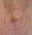
Skin tag. (WC)

Microscopic
Features:
- On a stalk / epithelium on three sides.
- Benign epidermis.
- Fibrous core.
- +/-Inflammation.
DDx:
- Pedunculated seborrheic keratosis - typically have epidermal hyperplasia, hyperkeratosis, horn cysts.[2]
- Regressing melanocytic lesions.
- Nevus lipomatosus superficialis - abundant adipocytes in the superficial dermis.
- Neurofibroma.
- Seborrheic keratosis - usu. horn cysts.
Images
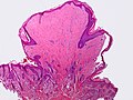
Skin tag. (WC)
www:
- Fibroepithelial polyp (dermatlas.med.jhmi.edu).[3]
- Fibroepithelial polyp (surgicalpathologyatlas.com).
- Inflamed fibroepithelial polyp (cdlib.org).[4]
Sign out
EYELID ("TAG"), RIGHT, REMOVAL:
- BENIGN FIBROEPITHELIAL POLYP.
SKIN ("SKIN TAG"), THIGH, REMOVAL:
- FIBROEPITHELIAL POLYP.
Micro
The sections show a fragment of skin with epithelium on three sides. The epithelium matures normally and is not hypertrophic. Orthokeratosis is present. The core of the lesion has fibrous tissue. There is no significant inflammation. No melanocytic nests are identified.
Hyperkeratosis
The sections show a fragment of skin with epithelium on three sides. The epidermis matures toward the surface and no significant basilar atypia is identified. A prominent granular layer is present. Mild hyperkeratosis is present. The core of the lesion consists of fibrous tissue. There is no significant inflammation. No melanocytic nests are identified.
Inflamed
The sections show a fragment of skin with epithelium on three sides. The epithelium matures and is acanthotic. Minimal parakeratosis is present. The core of the lesion consists of fibrous tissue with a mild lymphocyte-predominant dermal infiltrate. Rare siderophages are present.
There is mild basal nuclear enlargement. No significant nuclear atypia is apparent. The dermal-epidermal interface is well-demarcated. Rare basal mitotic activity is identified.
Epithelial-type
The sections show skin with epithelium on three sides, acanthosis and hyperkeratosis. No pseudohorn cysts are identified. A granular layer is present. Mild basal nuclear atypia is present. Mitotic activity is present but basal. Minimal parakeratosis is present. No melanocytic nests are identified. No solar elastosis is apparent in the limited dermis. No koilocytes are apparent.
See also
References
- ↑ Mitchell, Richard; Kumar, Vinay; Fausto, Nelson; Abbas, Abul K.; Aster, Jon (2011). Pocket Companion to Robbins & Cotran Pathologic Basis of Disease (8th ed.). Elsevier Saunders. pp. 596. ISBN 978-1416054542.
- ↑ Busam, Klaus J. (2009). Dermatopathology: A Volume in the Foundations in Diagnostic Pathology Series (1st ed.). Saunders. pp. 342. ISBN 978-0443066542.
- ↑ URL: http://dermatlas.med.jhmi.edu/derm/result.cfm?Diagnosis=1196583692. Accessed on: 1 September 2011.
- ↑ URL: http://dermatology-s10.cdlib.org/143/case_presentations/skintags/allegue.html. Accessed on: 9 January 2013.