Difference between revisions of "Ferruginous body"
Jump to navigation
Jump to search
(→Images) |
|||
| Line 22: | Line 22: | ||
===Images=== | ===Images=== | ||
<gallery> | <gallery> | ||
Image: Ferruginous bodies - BAL - r1 -- high mag.jpg | FB - high mag. | Image:Ferruginous_body.jpg | Ferruginous bodies. (WC) | ||
Image: Ferruginous bodies - BAL - r1 -- very high mag.jpg | FB - very high mag. | </gallery> | ||
====Cytology==== | |||
<gallery> | |||
Image: Ferruginous bodies - BAL - r1 -- high mag.jpg | FB - high mag. (WC) | |||
Image: Ferruginous bodies - BAL - r1 -- very high mag.jpg | FB - very high mag. (WC) | |||
Image: Ferruginous bodies - BAL - r2 -- very high mag.jpg | FB - very high mag. | Image: Ferruginous bodies - BAL - r2 -- very high mag.jpg | FB - very high mag. (WC) | ||
Image: Ferruginous bodies - BAL - r3 -- high mag.jpg | FB - high mag. | Image: Ferruginous bodies - BAL - r3 -- high mag.jpg | FB - high mag. (WC) | ||
Image: Ferruginous bodies - BAL - r3 -- very high mag.jpg | FB - very high mag. | Image: Ferruginous bodies - BAL - r3 -- very high mag.jpg | FB - very high mag. (WC) | ||
</gallery> | </gallery> | ||
<gallery> | |||
Image:Carcinoma asbestos body lung.jpg | Ferruginous body in carcinoma - cytology. (WC/Alex Brollo) | |||
</gallery> | |||
==Stains== | ==Stains== | ||
*Prussian blue +ve. | *Prussian blue +ve. | ||
Revision as of 04:59, 2 January 2016

Ferruginous body is a histopathologic finding in lung pathology that strongly suggest exposure to asbestos.
General
- Uncommon finding.
- Strongly suggestive of asbestos exposure.
Conditions associated with asbestos exposure (mnemonic PALM):[1]
Microscopic
Features:
- Segmented twirling baton with long slender fibre within.
- Black/brown crystal-like appearance.
DDx:
- Dirt - especially on H&E.
Images
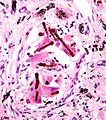
Ferruginous bodies. (WC)

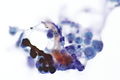
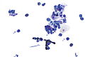
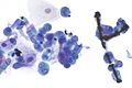
Cytology
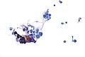
FB - high mag. (WC)
FB - very high mag. (WC)
FB - very high mag. (WC)
FB - high mag. (WC)
FB - very high mag. (WC)


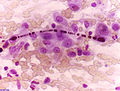
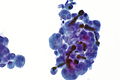
Ferruginous body in carcinoma - cytology. (WC/Alex Brollo)

Stains
- Prussian blue +ve.
See also
References
- ↑ Mitchell, Richard; Kumar, Vinay; Fausto, Nelson; Abbas, Abul K.; Aster, Jon (2011). Pocket Companion to Robbins & Cotran Pathologic Basis of Disease (8th ed.). Elsevier Saunders. pp. 375. ISBN 978-1416054542.