Eosinophilic esophagitis
Eosinophilic esophagitis, abbreviated EE, is relatively uncommon pathology of the esophagus with some similarities to gastroesophageal reflux disease (GERD).
General
- The current thinking is that it is a clinico-pathologic diagnosis.[1]
Clinical:
- Dysphagia[2] - classic presentation.
- Dyspepsia.
- Often mimics gastroesophageal reflux disease (GERD).[3]
Treatment:
- Avoid exacerbating antigens.
- Topical corticosteroids, e.g. fluticasone.
- Do not respond to proton pump inhibitors.
Biopsies:
- Should be taken from: upper, mid, lower and submitted in separate containers (eosinophilia present through-out-- to differentiate from GERD).
Associations:
- Atopy.[4]
- Celiac disease.[5]
- Oral antigens, i.e. particular foods.[3]
- Familial association.[3]
- Young ~ 35 years old.[6]
- Male > female (3:1).[6]
Gross/endoscopic
DDx (endoscopic):
Image
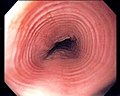
Trachealization of the esophagus. (WC)

Microscopic
Features:[4]
- Mucosa with "abundant eosinophils".
- Basal cell hyperplasia.
- Three cells thick or >15% of epithelial thickness.
- Papillae elongated.
- Papillae that reach into the top 1/3 of the epithelial layer - definition for GERD.[9]
Notes "abundant eosinophils":
- Criteria for number of eosinophils/area is highly variable; there is a 23X fold variation in published values and only 11% of studies actually define an area (most studies, embarassing for pathologists that understand this issue, only give the number of eosinophils per "HPF")![10]
- Interrater variability is low, i.e. good, if the procedure is standardized.[11]
- The most commonly reported cut points are 15, 20 and 24 eosinophils/HPF, without defining HPF.[10]
- The Foundation Series book[4] says: "> 20/HPF"; onlinepathology sees this definition as garbage, as "HPF" is not defined (see HPFitis).
- There is a consensus paper[12] that makes note of HPFitis... and then goes on to ignore to whole issue by defining EE as 15/HPF. It blows my mind that the people could be so will fully blind and that the idiotic reviewers didn't understand this.
- Most resident microscopes at the Toronto teaching hospitals have 22 mm eye pieces and have for their highest magnification objective a 40X. De facto, this means most people in Toronto are using the Liacouras et al. definition.[13]
- Eosinophils may be patchy.[14]
DDx:[15]
- Gastroesophageal reflux disease - no mid and proximal involvement.
- Infectious esophagitis.
- Eosinophilic gastroenteritis.
- Hypereosinophilic syndrome.
Images
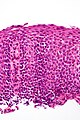
Eosinophilic esophagitis - very high mag. (WC)
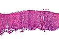
Eosinophilic esophagitis - high mag. (WC)


Sign out
ESOPHAGUS, DISTAL, BIOPSY:
- SQUAMOUS MUCOSA WITH BASAL CELL HYPERPLASIA, ABUNDANT INTRAEPITHELIAL EOSINOPHILS,
  EDEMA, AND PAPILLARY ELONGATION, SEE COMMENT.
- STAINS (PAS-D, GMS) NEGATIVE FOR MICROORGANISMS.
- NEGATIVE FOR INTESTINAL METAPLASIA.
- NEGATIVE FOR DYSPLASIA.

COMMENT:
There are approximately 65 eosinophils per 0.2376 mm*mm (1 HPF).

Literature valves show a large variation when defining eosinophilic esophagitis
and frequently use "HPF" as a measure of area, which is not a standardized measure.
[Am. J. Gastroenterol. 102 (10): 2300–13.]

Common cut-points are 15 eosinophils/HPF and 20 eosinophils/HPF, where HPF is
often undefined.

The above findings are suggestive of eosinophilic esophagitis in the proper
clinical context.
Patchy eosinophils
ESOPHAGUS (DISTAL), BIOPSY:
- SQUAMOUS MUCOSA WITH BASAL CELL HYPERPLASIA, INTRAEPITHELIAL EDEMA AND ONLY
  FOCALLY ABUNDANT INTRAEPITHELIAL EOSINOPHILS, SEE COMMENT.
- COLUMNAR EPITHELIUM WITH MODERATE CHRONIC INFLAMMATION, AND PANCREATIC
  ACINAR METAPLASIA.
- NEGATIVE FOR INTESTINAL METAPLASIA.
- NEGATIVE FOR DYSPLASIA AND NEGATIVE FOR MALIGNANCY.

COMMENT:
One high power field (field diameter 0.55 mm) has 25 eosinophils. The findings are
compatible with gastroesophageal reflux; however, eosinophilic esophagitis is also a
consideration. Clinical correlation is required.

Literature valves show a large variation when defining eosinophilic esophagitis
and frequently use "HPF" as a measure of area, which is not a standardized measure.
[Am. J. Gastroenterol. 102 (10): 2300 13.]

Common cut-points are 15 eosinophils/HPF and 20 eosinophils/HPF, where HPF is
often undefined.
Histology suggestive
ESOPHAGUS, BIOPSY:
- SQUAMOUS MUCOSA WITH MARKED BASAL CELL HYPERPLASIA, FOCALLY ABUNDANT
  INTRAEPITHELIAL EOSINOPHILS, EDEMA, AND PAPILLARY ELONGATION, SEE COMMENT.
- NEGATIVE FOR INTESTINAL METAPLASIA.
- NEGATIVE FOR DYSPLASIA.

COMMENT:
Focally, there are approximately 35 eosinophils per 0.2376 mm*mm (1 HPF).

The above findings raise the possibility of eosinophilic esophagitis; clinical
correlation is suggested. A re-biopsy including a portion of the proximal esophagus
could be considered.
See also
References
- ↑ 1.0 1.1 Genevay, M.; Rubbia-Brandt, L.; Rougemont, AL. (Jun 2010). "Do eosinophil numbers differentiate eosinophilic esophagitis from gastroesophageal reflux disease?". Arch Pathol Lab Med 134 (6): 815-25. doi:10.1043/1543-2165-134.6.815. PMID 20524860. http://www.archivesofpathology.org/doi/full/10.1043/1543-2165-134.6.815.
- ↑ URL: http://www.medicinenet.com/eosinophilic_esophagitis/page2.htm#tocc. Accessed on: 1 December 2009.
- ↑ 3.0 3.1 3.2 Rothenberg, ME. (Oct 2009). "Biology and treatment of eosinophilic esophagitis.". Gastroenterology 137 (4): 1238-49. doi:10.1053/j.gastro.2009.07.007. PMID 19596009.
- ↑ 4.0 4.1 4.2 Iacobuzio-Donahue, Christine A.; Montgomery, Elizabeth A. (2005). Gastrointestinal and Liver Pathology: A Volume in the Foundations in Diagnostic Pathology Series (1st ed.). Churchill Livingstone. pp. 19. ISBN 978-0443066573.
- ↑ Leslie C, Mews C, Charles A, Ravikumara M (April 2010). "Celiac disease and eosinophilic esophagitis: a true association". J. Pediatr. Gastroenterol. Nutr. 50 (4): 397–9. doi:10.1097/MPG.0b013e3181a70af4. PMID 19841598.
- ↑ 6.0 6.1 Dellon, ES.; Erichsen, R.; Pedersen, L.; Shaheen, NJ.; Baron, JA.; Sørensen, HT.; Vyberg, M. (Jan 2013). "Development and validation of a registry-based definition of eosinophilic esophagitis in Denmark.". World J Gastroenterol 19 (4): 503-10. doi:10.3748/wjg.v19.i4.503. PMID 23382628.
- ↑ Al-Hussaini, AA.; Semaan, T.; El Hag, IA.. "Esophageal trachealization: a feature of eosinophilic esophagitis.". Saudi J Gastroenterol 15 (3): 193-5. doi:10.4103/1319-3767.54747. PMID 19636182.
- ↑ URL: http://www.ajronline.org/cgi/reprint/164/4/900.pdf. Accessed on: 4 October 2010.
- ↑ Cotran, Ramzi S.; Kumar, Vinay; Fausto, Nelson; Nelso Fausto; Robbins, Stanley L.; Abbas, Abul K. (2005). Robbins and Cotran pathologic basis of disease (7th ed.). St. Louis, Mo: Elsevier Saunders. pp. 804. ISBN 0-7216-0187-1.
- ↑ 10.0 10.1 Dellon ES, Aderoju A, Woosley JT, Sandler RS, Shaheen NJ (October 2007). "Variability in diagnostic criteria for eosinophilic esophagitis: a systematic review". Am. J. Gastroenterol. 102 (10): 2300–13. doi:10.1111/j.1572-0241.2007.01396.x. PMID 17617209.
- ↑ Dellon, ES.; Fritchie, KJ.; Rubinas, TC.; Woosley, JT.; Shaheen, NJ. (Jul 2010). "Inter- and intraobserver reliability and validation of a new method for determination of eosinophil counts in patients with esophageal eosinophilia.". Dig Dis Sci 55 (7): 1940-9. doi:10.1007/s10620-009-1005-z. PMID 19830560.
- ↑ Furuta GT, Liacouras CA, Collins MH, et al. (October 2007). "Eosinophilic esophagitis in children and adults: a systematic review and consensus recommendations for diagnosis and treatment". Gastroenterology 133 (4): 1342–63. doi:10.1053/j.gastro.2007.08.017. PMID 17919504.
- ↑ Liacouras CA, Spergel JM, Ruchelli E, et al. (December 2005). "Eosinophilic esophagitis: a 10-year experience in 381 children". Clin. Gastroenterol. Hepatol. 3 (12): 1198–206. PMID 16361045.
- ↑ Saffari, H.; Peterson, KA.; Fang, JC.; Teman, C.; Gleich, GJ.; Pease, LF. (Sep 2012). "Patchy eosinophil distributions in an esophagectomy specimen from a patient with eosinophilic esophagitis: Implications for endoscopic biopsy.". J Allergy Clin Immunol 130 (3): 798-800. doi:10.1016/j.jaci.2012.03.009. PMID 22502795.
- ↑ Odze, Robert D.; Goldblum, John R. (2009). Surgical pathology of the GI tract, liver, biliary tract and pancreas (2nd ed.). Saunders. pp. 244. ISBN 978-1416040590.