Difference between revisions of "Dural tail sign"
Jump to navigation
Jump to search
(create) |
Jensflorian (talk | contribs) |
||
| (3 intermediate revisions by one other user not shown) | |||
| Line 1: | Line 1: | ||
'''Dural tail sign''' a finding in neuroradiology associated with [[meningioma]],<ref name=pmid2120998>{{Cite journal | last1 = Aoki | first1 = S. | last2 = Sasaki | first2 = Y. | last3 = Machida | first3 = T. | last4 = Tanioka | first4 = H. | title = Contrast-enhanced MR images in patients with meningioma: importance of enhancement of the dura adjacent to the tumor. | journal = AJNR Am J Neuroradiol | volume = 11 | issue = 5 | pages = 935-8 | month = | year = | doi = | PMID = 2120998 }}</ref> but also described in other tumours. | '''Dural tail sign''', abbreviated '''DTS''', is a finding in neuroradiology associated with [[meningioma]],<ref name=pmid2120998>{{Cite journal | last1 = Aoki | first1 = S. | last2 = Sasaki | first2 = Y. | last3 = Machida | first3 = T. | last4 = Tanioka | first4 = H. | title = Contrast-enhanced MR images in patients with meningioma: importance of enhancement of the dura adjacent to the tumor. | journal = AJNR Am J Neuroradiol | volume = 11 | issue = 5 | pages = 935-8 | month = | year = | doi = | PMID = 2120998 }}</ref> but also described in other tumours.<ref name=pmid21161034>{{Cite journal | last1 = Sotoudeh | first1 = H. | last2 = Yazdi | first2 = HR. | title = A review on dural tail sign. | journal = World J Radiol | volume = 2 | issue = 5 | pages = 188-92 | month = May | year = 2010 | doi = 10.4329/wjr.v2.i5.188 | PMID = 21161034 }}</ref> | ||
It is seen on MRI T1 images as a thickening with enhancement adjacent to a (mass) lesion.<ref name=pmid21161034/> | |||
==Image== | |||
<gallery> | |||
File:Keilbeinmeningeom_MRT_T1KMsag.png | Dural tail sign in a meningioma, T1 sagittal plane. | |||
</gallery> | |||
==See also== | ==See also== | ||
Latest revision as of 08:16, 9 December 2015
Dural tail sign, abbreviated DTS, is a finding in neuroradiology associated with meningioma,[1] but also described in other tumours.[2]
It is seen on MRI T1 images as a thickening with enhancement adjacent to a (mass) lesion.[2]
Image
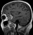
Dural tail sign in a meningioma, T1 sagittal plane.

See also
References
- ↑ Aoki, S.; Sasaki, Y.; Machida, T.; Tanioka, H.. "Contrast-enhanced MR images in patients with meningioma: importance of enhancement of the dura adjacent to the tumor.". AJNR Am J Neuroradiol 11 (5): 935-8. PMID 2120998.
- ↑ 2.0 2.1 Sotoudeh, H.; Yazdi, HR. (May 2010). "A review on dural tail sign.". World J Radiol 2 (5): 188-92. doi:10.4329/wjr.v2.i5.188. PMID 21161034.