Difference between revisions of "Diffuse tenosynovial giant-cell tumour"
Jump to navigation
Jump to search
| Line 36: | Line 36: | ||
==General== | ==General== | ||
* | *Usually benign. | ||
**Occasionally malignant.<ref name=pmid22827766>{{Cite journal | last1 = Kondo | first1 = R. | last2 = Akiba | first2 = J. | last3 = Hiraoka | first3 = K. | last4 = Hisaoka | first4 = M. | last5 = Hashimoto | first5 = H. | last6 = Kage | first6 = M. | last7 = Yano | first7 = H. | title = Malignant diffuse-type tenosynovial giant cell tumor of the buttock. | journal = Pathol Int | volume = 62 | issue = 8 | pages = 559-64 | month = Aug | year = 2012 | doi = 10.1111/j.1440-1827.2012.02838.x | PMID = 22827766 }}</ref><ref name=pmid18301053>{{Cite journal | last1 = Li | first1 = CF. | last2 = Wang | first2 = JW. | last3 = Huang | first3 = WW. | last4 = Hou | first4 = CC. | last5 = Chou | first5 = SC. | last6 = Eng | first6 = HL. | last7 = Lin | first7 = CN. | last8 = Yu | first8 = SC. | last9 = Huang | first9 = HY. | title = Malignant diffuse-type tenosynovial giant cell tumors: a series of 7 cases comparing with 24 benign lesions with review of the literature. | journal = Am J Surg Pathol | volume = 32 | issue = 4 | pages = 587-99 | month = Apr | year = 2008 | doi = 10.1097/PAS.0b013e318158428f | PMID = 18301053 }}</ref><ref name=pmid10757395>{{Cite journal | last1 = Somerhausen | first1 = NS. | last2 = Fletcher | first2 = CD. | title = Diffuse-type giant cell tumor: clinicopathologic and immunohistochemical analysis of 50 cases with extraarticular disease. | journal = Am J Surg Pathol | volume = 24 | issue = 4 | pages = 479-92 | month = Apr | year = 2000 | doi = | PMID = 10757395 }}</ref> | **Occasionally malignant.<ref name=pmid22827766>{{Cite journal | last1 = Kondo | first1 = R. | last2 = Akiba | first2 = J. | last3 = Hiraoka | first3 = K. | last4 = Hisaoka | first4 = M. | last5 = Hashimoto | first5 = H. | last6 = Kage | first6 = M. | last7 = Yano | first7 = H. | title = Malignant diffuse-type tenosynovial giant cell tumor of the buttock. | journal = Pathol Int | volume = 62 | issue = 8 | pages = 559-64 | month = Aug | year = 2012 | doi = 10.1111/j.1440-1827.2012.02838.x | PMID = 22827766 }}</ref><ref name=pmid18301053>{{Cite journal | last1 = Li | first1 = CF. | last2 = Wang | first2 = JW. | last3 = Huang | first3 = WW. | last4 = Hou | first4 = CC. | last5 = Chou | first5 = SC. | last6 = Eng | first6 = HL. | last7 = Lin | first7 = CN. | last8 = Yu | first8 = SC. | last9 = Huang | first9 = HY. | title = Malignant diffuse-type tenosynovial giant cell tumors: a series of 7 cases comparing with 24 benign lesions with review of the literature. | journal = Am J Surg Pathol | volume = 32 | issue = 4 | pages = 587-99 | month = Apr | year = 2008 | doi = 10.1097/PAS.0b013e318158428f | PMID = 18301053 }}</ref><ref name=pmid10757395>{{Cite journal | last1 = Somerhausen | first1 = NS. | last2 = Fletcher | first2 = CD. | title = Diffuse-type giant cell tumor: clinicopathologic and immunohistochemical analysis of 50 cases with extraarticular disease. | journal = Am J Surg Pathol | volume = 24 | issue = 4 | pages = 479-92 | month = Apr | year = 2000 | doi = | PMID = 10757395 }}</ref> | ||
* | *Can be thought of as the large joint version of [[giant cell tumour of the tendon sheath]].<ref name=Ref_DCHH341>{{Ref DCHH|341}}</ref> | ||
===Classification=== | ===Classification=== | ||
Revision as of 18:53, 5 May 2014
| Diffuse tenosynovial giant-cell tumour | |
|---|---|
| Diagnosis in short | |
 Diffuse tenosynovial giant-cell tumour. H&E stain. | |
|
| |
| Synonyms | pigmented villonodular synovitis (PVNS) - old term |
|
| |
| LM | nodules composed of cells with abundant cytoplasm & pale nuclei, multinucleated giant cells, hemosiderin-laden macrophages, foam cells |
| Subtypes | generalized, localized (articular, extra-articular) |
| LM DDx | giant cell lesions, others |
| Gross | pigmented, articular or extra-articular |
| Site | large joints - esp. knee or hip; occ. extra-articular |
|
| |
| Prevalence | uncommon |
| Prognosis | usually benign |
Diffuse tenosynovial giant-cell tumour is relatively common mostly benign chondro-osseous tumour of the large joints.
It is also known as tenosynovial giant-cell tumour, diffuse type. Previously, it was known as pigmented villonodular synovitis, abbreviated PVNS.[1]
General
- Usually benign.
- Can be thought of as the large joint version of giant cell tumour of the tendon sheath.[5]
Classification
Subclassified - clinical:[6]
- Generalized PVNS.
- Localized PVNS.[7]
- Articular.
- Extra-articular.
Gross
Note:
- Localized form - classically fat pad inferior to patella.[9]
Microscopic
Features:[10]
- Subsynovial nodules composed of cells with:
- Abundant cytoplasm.
- Pale nuclei.
- Multinucleated giant cells.
- Hemosiderin-laden macrophages.
- Foam cells.
DDx - general for the site:[11]
- Synovial chondromatosis.
- Gout.
- Pseudogout.
- Storage disorders.
- Granulomatous inflammation.
- Degenerative changes (osteoarthritis).
- Rheumatic disease.
Images
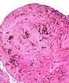
PVNS - low mag. (WC)
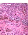
PVNS - high mag. (WC)

www:
- PVNS - several images (upmc.edu).
- PVNS (webpathology.com).
- Localized nodular synovitis (nih.gov).[9]
Molecular
- Clonal - overexpresses CSF1.[12]
Sign out
RIGHT FEMORAL HEAD AND JOINT CAPSULE, EXCISION:
- DEGENERATIVE JOINT DISEASE.
- DIFFUSE TENOSYNOVIAL GIANT-CELL TUMOUR (PIGMENTED VILLONODULAR SYNOVITIS).
Micro
The soft tissue sections show nodules with abundant hemosiderin-laden macrophages and multinucleated giant cells. Nuclear atypia is not identified. Mitotic activity is not apparent.
See also
References
- ↑ Kumar, Vinay; Abbas, Abul K.; Fausto, Nelson; Aster, Jon (2009). Robbins and Cotran pathologic basis of disease (8th ed.). Elsevier Saunders. pp. 1247. ISBN 978-1416031215.
- ↑ Kondo, R.; Akiba, J.; Hiraoka, K.; Hisaoka, M.; Hashimoto, H.; Kage, M.; Yano, H. (Aug 2012). "Malignant diffuse-type tenosynovial giant cell tumor of the buttock.". Pathol Int 62 (8): 559-64. doi:10.1111/j.1440-1827.2012.02838.x. PMID 22827766.
- ↑ Li, CF.; Wang, JW.; Huang, WW.; Hou, CC.; Chou, SC.; Eng, HL.; Lin, CN.; Yu, SC. et al. (Apr 2008). "Malignant diffuse-type tenosynovial giant cell tumors: a series of 7 cases comparing with 24 benign lesions with review of the literature.". Am J Surg Pathol 32 (4): 587-99. doi:10.1097/PAS.0b013e318158428f. PMID 18301053.
- ↑ 4.0 4.1 Somerhausen, NS.; Fletcher, CD. (Apr 2000). "Diffuse-type giant cell tumor: clinicopathologic and immunohistochemical analysis of 50 cases with extraarticular disease.". Am J Surg Pathol 24 (4): 479-92. PMID 10757395.
- ↑ Tadrous, Paul.J. Diagnostic Criteria Handbook in Histopathology: A Surgical Pathology Vade Mecum (1st ed.). Wiley. pp. 341. ISBN 978-0470519035.
- ↑ Perka, C.; Labs, K.; Zippel, H.; Buttgereit, F. (Feb 2000). "Localized pigmented villonodular synovitis of the knee joint: neoplasm or reactive granuloma? A review of 18 cases.". Rheumatology (Oxford) 39 (2): 172-8. PMID 10725067.
- ↑ Huang, GS.; Lee, CH.; Chan, WP.; Chen, CY.; Yu, JS.; Resnick, D. (Aug 2003). "Localized nodular synovitis of the knee: MR imaging appearance and clinical correlates in 21 patients.". AJR Am J Roentgenol 181 (2): 539-43. doi:10.2214/ajr.181.2.1810539. PMID 12876042.
- ↑ Frassica, FJ.; Bhimani, MA.; McCarthy, EF.; Wenz, J. (Oct 1999). "Pigmented villonodular synovitis of the hip and knee.". Am Fam Physician 60 (5): 1404-10; discussion 1415. PMID 10524485.
- ↑ 9.0 9.1 Park, JH.; Ro, KH.; Lee, DH. (May 2013). "Localized nodular synovitis of the infrapatellar fat pad.". Indian J Orthop 47 (3): 313-6. doi:10.4103/0019-5413.111514. PMID 23798766.
- ↑ URL: http://www.wheelessonline.com/ortho/pigmented_villonodular_synovitis.
- ↑ Krenn, V.; Morawietz, L.; König, A.; Haeupl, T. (Nov 2006). "[Differential diagnosis of chronic synovitis].". Pathologe 27 (6): 402-8. doi:10.1007/s00292-006-0866-6. PMID 17031677.
- ↑ Lucas, DR. (Aug 2012). "Tenosynovial giant cell tumor: case report and review.". Arch Pathol Lab Med 136 (8): 901-6. doi:10.5858/arpa.2012-0165-CR. PMID 22849738.