Dermal scar
Jump to navigation
Jump to search
| Dermal scar | |
|---|---|
| Diagnosis in short | |
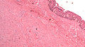
 Dermal scar. H&E stain. | |
|
| |
| LM | dense collagen - fibers run parallel to the DE junction, loss of dermal papilla, loss of adnexal structures, thin-wall blood vessels |
| LM DDx | malignant melanoma desmoplastic-neurotropic type, dermatofibroma, desmoplastic Spitz nevus, sclerosing blue nevus |
| Stains | S-100 -ve (mostly) |
| Site | skin |
|
| |
| Clinical history | trauma, previous excision or biopsy |
| Prevalence | common |
| Prognosis | benign |
| Dermal scar | |
|---|---|
| External resources | |
| EHVSC | 10187 (Dermal scar adjacent to a basal cell carcinoma) |
| Wikipedia | Scar |
Dermal scar, also simply scar, is commonly seen in dermatopathology. It is also known a cicatrix.
General
- Previous surgery, biopsy, trauma.
Microscopic
Features:
- Loss of dermal papilla.
- Dense collagen - fibers run parallel to the dermal-epidermal (DE) junction[1] - key feature.
- Loss of adnexal structures.
Other feature:
- Thin-walled blood vessels.
- Described as running perpendicular to the surface[1] - this may not be apparent.
Note:
- There should not be any nuclear hyperchromasia or pleomorphism.[2]
DDx:
- Malignant melanoma, desmoplastic-neurotropic type - nuclear pleomorphism and/or hyperchromasia; may be focal.[2]
- Dermatofibroma.
- Desmoplastic Spitz nevus.
- Sclerosing blue nevus.
Image
Scar. (WC)
IHC
- S100 focal/scattered +ve.
- Desmoplastic melanoma strong +ve.
- HMB-45 -ve.
- Sclerosing blue nevus +ve.
Sign out
SKIN, LOWER MID BACK, RE-EXCISION:
- DERMAL SCAR.
- SOLAR ELASTOSIS.
Micro
The sections show skin with a dermis with dense collagen fibres that run parallel to the skin surface without adnexal structures. The overlying dermal-epidermis interface lacks the typical undulation.
See also
References
- ↑ 1.0 1.1 Busam, Klaus J. (2009). Dermatopathology: A Volume in the Foundations in Diagnostic Pathology Series (1st ed.). Saunders. pp. 499. ISBN 978-0443066542.
- ↑ 2.0 2.1 Busam, Klaus J. (2009). Dermatopathology: A Volume in the Foundations in Diagnostic Pathology Series (1st ed.). Saunders. pp. 479. ISBN 978-0443066542.