Difference between revisions of "Cystic medial degeneration"
Jump to navigation
Jump to search
| Line 13: | Line 13: | ||
*Disruption of the elastic lamina (seen on elastic trichrome stain). | *Disruption of the elastic lamina (seen on elastic trichrome stain). | ||
*+/-Focal necrosis. | *+/-Focal necrosis. | ||
DDx: | |||
*[[Aortic dissection]] without apparent underlying pathology. | |||
===Images=== | ===Images=== | ||
| Line 30: | Line 33: | ||
==See also== | ==See also== | ||
*[[Vascular disease]]. | *[[Vascular disease]]. | ||
*[[Aortic dissection]]. | |||
==References== | ==References== | ||
Revision as of 16:59, 16 May 2014
Cystic medial degeneration (abbreviated CMD), also cystic medial necrosis,[1] is vascular pathology of the large blood vessels. It is suggestive of an underlying connective tissue disorder.
General
- Nonspecific finding - may be seen in a number of conditions.
- "Medial" refers to tunica media the middle (muscle) layer of an artery.
Note about cystic medial necrosis:
- Often not cystic and not necrotic.
Microscopic
- Basophilic ground substance in the media (seen on Movat's stain).
- Disruption of the elastic lamina (seen on elastic trichrome stain).
- +/-Focal necrosis.
DDx:
- Aortic dissection without apparent underlying pathology.
Images
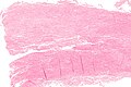
Cystic medial degeneration - low mag. (WC/Nephron)
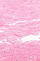
Cystic medial degeneration - high mag. (WC/Nephron)
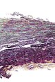
Cystic medial degeneration - movat - low mag. (WC/Nephron)
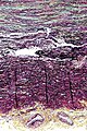
Cystic medial degeneration - movat - intermed. mag. (WC/Nephron)


www:
Stains
- Elastin stains (e.g. elastic trichrome stain) - disruption of the elastic lamina.
- Movat's stain - basophilic ground substance in the media.
See also
References
- ↑ URL: http://emedicine.medscape.com/article/756835-overview. Accessed on: 12 August 2010.
- ↑ URL: http://emedicine.medscape.com/article/756835-overview. Accessed on: 12 August 2010.
- ↑ Ha HI, Seo JB, Lee SH, et al. (2007). "Imaging of Marfan syndrome: multisystemic manifestations". Radiographics 27 (4): 989–1004. doi:10.1148/rg.274065171. PMID 17620463. http://radiographics.rsna.org/content/27/4/989.full.