Difference between revisions of "Cutaneous calcinosis"
Jump to navigation
Jump to search
(+cat.) |
(split out) |
||
| Line 1: | Line 1: | ||
# | {{ Infobox diagnosis | ||
| Name = {{PAGENAME}} | |||
| Image = Calcinosis cutis -- low mag.jpg | |||
| Width = | |||
| Caption = Cutaneous calcinosis. [[H&E stain]]. | |||
| Synonyms = | |||
| Micro = dermal calcification - usu. well-circumscribed | |||
| Subtypes = | |||
| LMDDx = | |||
| Stains = | |||
| IHC = | |||
| EM = | |||
| Molecular = | |||
| IF = | |||
| Gross = firm nodule | |||
| Grossing = | |||
| Site = [[skin]], [[scrotum]] | |||
| Assdx = | |||
| Syndromes = | |||
| Clinicalhx = +/-trauma at the site | |||
| Signs = firm nodule | |||
| Symptoms = | |||
| Prevalence = uncommon | |||
| Bloodwork = | |||
| Rads = | |||
| Endoscopy = | |||
| Prognosis = benign | |||
| Other = | |||
| ClinDDx = | |||
| Tx = excision | |||
}} | |||
'''Cutaneous calcinosis''', also '''calcinosis cutis''', is calcification of the [[skin]]. It is benign in itself; however, the underlying cause not be. | |||
==General== | |||
*Benign in itself; underlying cause may not be benign. | |||
*May be a [[scrotum|scrotal]] lesion - known as ''scrotal calcinosis''.<ref name=pmid20178701>{{Cite journal | last1 = Dubey | first1 = S. | last2 = Sharma | first2 = R. | last3 = Maheshwari | first3 = V. | title = Scrotal calcinosis: idiopathic or dystrophic? | journal = Dermatol Online J | volume = 16 | issue = 2 | pages = 5 | month = | year = 2010 | doi = | PMID = 20178701 }}</ref> | |||
Subtypes:<ref name=emed>URL: [http://emedicine.medscape.com/article/1103137-overview http://emedicine.medscape.com/article/1103137-overview]. Accessed on: 21 September 2011.</ref> | |||
#Dystrophic - due to death of cells; may be related to a tumour. | |||
#Metastatic - due to chronic renal failure; hyperkalemia; paraneoplastic phenomenon. | |||
#Iatrogenic - post surgical. | |||
#Idiopathic. | |||
==Gross== | |||
*Firm nodule. | |||
==Microscopic== | |||
Features: | |||
*Dermal calcification: | |||
**Acellular purple blobs on [[H&E]]. | |||
***+/-Artefactual tearing of surrounding tissue due to processing (cutting). | |||
***+/-Small artefactual lines ~1-2 micrometers due to processing (cutting). | |||
***+/-Greyish rim of paucicellular material. | |||
**Usu. well-circumscribed. | |||
***May be surrounded by a palisading granuloma & [[giant cell]]s. | |||
===Images=== | |||
<gallery> | |||
Image: Calcinosis cutis -- very low mag.jpg | CC - very low mag. | |||
Image: Calcinosis cutis -- low mag.jpg | CC - low mag. | |||
Image: Calcinosis cutis -- intermed mag.jpg | CC - intermed. mag. | |||
</gallery> | |||
www: | |||
*[http://www.dermatopathonline.com/calcinosis%20cutis2.html Calcinosis cutis (dermatopathonline.com)]. | |||
==Sign out== | |||
<pre> | |||
SKIN AND SUBCUTANEOUS LESION, LEFT HIP, EXCISION: | |||
- SUBCUTANEOUS CALCIFICATION SURROUNDED BY BENIGN FIBROUS TISSUE. | |||
- DERMAL SCAR. | |||
- NEGATIVE FOR MALIGNANCY. | |||
</pre> | |||
<pre> | |||
SUBCUTANEOUS MASS, OVER BURSA OF ELBOW, EXCISION: | |||
- CALCINOSIS CUTIS. | |||
</pre> | |||
===Micro=== | |||
The sections show subcutaneous calcifications surrounded by macrophages and giant cells. No nuclear atypia is apparent. The overlying skin is unremarkable. | |||
==See also== | |||
*[[Non-malignant skin disease]]. | |||
==References== | |||
{{Reflist|2}} | |||
[[Category:Diagnosis]] | [[Category:Diagnosis]] | ||
[[Category:Non-malignant skin disease]] | |||
Revision as of 02:58, 2 January 2014
| Cutaneous calcinosis | |
|---|---|
| Diagnosis in short | |
 Cutaneous calcinosis. H&E stain. | |
|
| |
| LM | dermal calcification - usu. well-circumscribed |
| Gross | firm nodule |
| Site | skin, scrotum |
|
| |
| Clinical history | +/-trauma at the site |
| Signs | firm nodule |
| Prevalence | uncommon |
| Prognosis | benign |
| Treatment | excision |
Cutaneous calcinosis, also calcinosis cutis, is calcification of the skin. It is benign in itself; however, the underlying cause not be.
General
- Benign in itself; underlying cause may not be benign.
- May be a scrotal lesion - known as scrotal calcinosis.[1]
Subtypes:[2]
- Dystrophic - due to death of cells; may be related to a tumour.
- Metastatic - due to chronic renal failure; hyperkalemia; paraneoplastic phenomenon.
- Iatrogenic - post surgical.
- Idiopathic.
Gross
- Firm nodule.
Microscopic
Features:
- Dermal calcification:
- Acellular purple blobs on H&E.
- +/-Artefactual tearing of surrounding tissue due to processing (cutting).
- +/-Small artefactual lines ~1-2 micrometers due to processing (cutting).
- +/-Greyish rim of paucicellular material.
- Usu. well-circumscribed.
- May be surrounded by a palisading granuloma & giant cells.
- Acellular purple blobs on H&E.
Images
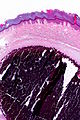
CC - very low mag.
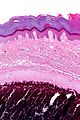
CC - low mag.
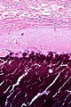
CC - intermed. mag.


www:
Sign out
SKIN AND SUBCUTANEOUS LESION, LEFT HIP, EXCISION:
- SUBCUTANEOUS CALCIFICATION SURROUNDED BY BENIGN FIBROUS TISSUE.
- DERMAL SCAR.
- NEGATIVE FOR MALIGNANCY.
SUBCUTANEOUS MASS, OVER BURSA OF ELBOW, EXCISION:
- CALCINOSIS CUTIS.
Micro
The sections show subcutaneous calcifications surrounded by macrophages and giant cells. No nuclear atypia is apparent. The overlying skin is unremarkable.
See also
References
- ↑ Dubey, S.; Sharma, R.; Maheshwari, V. (2010). "Scrotal calcinosis: idiopathic or dystrophic?". Dermatol Online J 16 (2): 5. PMID 20178701.
- ↑ URL: http://emedicine.medscape.com/article/1103137-overview. Accessed on: 21 September 2011.