Cutaneous T-cell lymphoma
Jump to navigation
Jump to search
The printable version is no longer supported and may have rendering errors. Please update your browser bookmarks and please use the default browser print function instead.
Cutaneous T-cell lymphoma, abbreviated CTCL, is a malignant lymphoid neoplasm that arises in the skin.
General
- Mycosis fungoides - is a subtype (???).
- CTCL is more common than cutaneous B-cell lymphoma (CBCL).[1][2]
Stages - like Kaposi sarcoma:
- Patch.
- Plaque.
- Nodular.
Microscopic
- Atypical lymphocytes:
- Have folded "cerebriform" nuclei; Sezary-Lutzner cells.[3]
- Grouping:
- Nests in the epidermis - known as "Pautrier microabscesses".
- Single lymphocytes in epidermis - without accompanying edema.
- Short linear arrays of lymphocytes along the basal layer of the epidermis; "epidermotropism".[3]
DDx:
Images
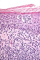
CTCL - very high mag. (WC/Nephron)
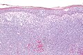
CTCL - intermed. mag. (WC/Nephron)


www:
IHC
Key stain:
- CD4 +ve.[4]
Other stains:
- CD3 +ve.
- CD8 -ve.
- CD20 -ve (to r/o significant B cell population).
- CD30 -ve.
- CD5 +ve.
- CD7 -ve (often lost first in T cell lymphomas).
- Ki-67 high.
- CD56 -ve.
See also
References
- ↑ URL: http://emedicine.medscape.com/article/1099540-overview. Accessed on: 24 August 2010.
- ↑ URL: http://emedicine.medscape.com/article/1098342-overview. Accessed on: 24 August 2010.
- ↑ 3.0 3.1 Klatt, Edward C. (2006). Robbins and Cotran Atlas of Pathology (1st ed.). Saunders. pp. 385. ISBN 978-1416002741.
- ↑ Kumar, Vinay; Abbas, Abul K.; Fausto, Nelson; Aster, Jon (2009). Robbins and Cotran pathologic basis of disease (8th ed.). Elsevier Saunders. pp. 1185. ISBN 978-1416031215.