Crystals in body fluids
This article deals with crystals in body fluids.
Crystals
Joint crystals
Types:[1]
- Gout = needle-shaped, negatively birefringent, yellow when aligned.
- Pseudogout = rhomboid-shaped, positively birefringent, blue when aligned.
Notes:
- Pseudogout also known as CPPD = calcium pyrophosphate dehydrogenase.
- Memory device: ABC+ = aligned blue is calcium & cuboid - positively birefringent.
Urine crystals
Types - morphology:
- Envelope shape (calcium oxalate).
- Diamond shape (uric acid).
- Coffin-lid shape (struvite).
- Hexagonal shape (cysteine).
Notes:
- Memory devices:
- Diamonds are see-through; ergo, uric acid stones not seen on KUB.
- Calcium oxalate = envelope, uric acid = diamond.
- Uric acid crystals: usually dissolve in formalin... but do not dissolve in alcohol.[2]
- Calcium oxalate crystals are seen in the context of ethylene glycol poisoning.[3]
Diseases
Gout
General
- Classically afflicts the big toe - known as podagra.
Etiology:[4]
- Overproduction of uric acid ~ 10% of cases.
- Reduced excretion of uric acid ~ 90% of cases.
Gross/radiology
- Radiologically may mimic anconeus epitrochlearis muscle.[7]
Microscopic
Features:[8]
- Tophi (advanced)
- Reactive granulomatous inflammation.
- Surrounds fluffy (cotton candy-like) material.
- Fibrotic synovium.
- Aggregates of urate crystals.
- Reactive granulomatous inflammation.
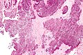
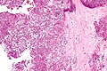
Images:
- Gouty tophus - A. xray, B. Diff-Quick, C. Pap smear, D. polarized light, E. H&E (archivesofpathology.org).
- Gouty tophus - low mag. (WC).
- Gouty tophus - high mag. (WC).
- Gout - several images (upmc.edu).
Pseudogout
- Chondrocalcinosis redirects here.
General
- Classically found in the knee.
Radiology
- Similar to osteoarthritis - joint space narrowing & subchondral sclerosis.[10]
Findings suggestive of CPPD:[10]
- Subchondral cysts (large) - important.
- Intra-articular bodies, several.
- Increased narrowing of patellofemoral joint.
Microscopic
Features:
- Crystals with a rhomboid-shape.
- Positively birefringent, blue when aligned.
Notes:
- Memory device: ABC+ = aligned blue is calcium & cuboid - positively birefringent.
Images
Pseudogout - intermed. mag. (WC)
Pseudogout - high mag. (WC)


www:
See also
References
- ↑ Yeung, J.C.; Leonard, Blair J. N. (2005). The Toronto Notes 2005 - Review for the MCCQE and Comprehensive Medical Reference (2005 ed.). The Toronto Notes Inc. for Medical Students Inc.. pp. RH6. ISBN 978-0968592854.
- ↑ WG. 8 January 2010.
- ↑ Saukko, Pekka; Knight, Bernard (2004). Knight's Forensic Pathology (3rd ed.). A Hodder Arnold Publication. pp. 589. ISBN 978-0340760444.
- ↑ Mitchell, Richard; Kumar, Vinay; Fausto, Nelson; Abbas, Abul K.; Aster, Jon (2011). Pocket Companion to Robbins & Cotran Pathologic Basis of Disease (8th ed.). Elsevier Saunders. pp. 634. ISBN 978-1416054542.
- ↑ Online 'Mendelian Inheritance in Man' (OMIM) 607096
- ↑ Tin, A.; Woodward, OM.; Kao, WH.; Liu, CT.; Lu, X.; Nalls, MA.; Shriner, D.; Semmo, M. et al. (Oct 2011). "Genome-wide association study for serum urate concentrations and gout among African Americans identifies genomic risk loci and a novel URAT1 loss-of-function allele.". Hum Mol Genet 20 (20): 4056-68. doi:10.1093/hmg/ddr307. PMID 21768215.
- ↑ URL: http://radiology.casereports.net/index.php/rcr/article/viewArticle/57/213. Accessed on: 7 August 2011.
- ↑ URL: http://pathologyoutlines.com/joints.html#gout. Accessed on: 5 August 2011.
- ↑ URL: http://www.ncbi.nlm.nih.gov/pubmedhealth/PMH0001458/. Accessed on: 28 October 2011.
- ↑ 10.0 10.1 URL: http://www.learningradiology.com/archives04/COW%20102-CPPD/cppdcorrectpage.htm. Accessed on: 9 October 2013.
- ↑ Dieppe, P.; Swan, A. (May 1999). "Identification of crystals in synovial fluid.". Ann Rheum Dis 58 (5): 261-3. PMC 1752883. PMID 10225806. https://www.ncbi.nlm.nih.gov/pmc/articles/PMC1752883/.