Difference between revisions of "Crystals in body fluids"
Jump to navigation
Jump to search
(→Gout: split-out) |
|||
| Line 27: | Line 27: | ||
=Diseases= | =Diseases= | ||
==Gout== | ==Gout== | ||
{{Main|Gout}} | |||
==Pseudogout== | ==Pseudogout== | ||
Revision as of 02:21, 4 February 2014
This article deals with crystals in body fluids.
Crystals
Joint crystals
Types:[1]
- Gout = needle-shaped, negatively birefringent, yellow when aligned.
- Pseudogout = rhomboid-shaped, positively birefringent, blue when aligned.
Notes:
- Pseudogout also known as CPPD = calcium pyrophosphate dehydrogenase.
- Memory device: ABC+ = aligned blue is calcium & cuboid - positively birefringent.
Urine crystals
Types - morphology:
- Envelope shape (calcium oxalate).
- Diamond shape (uric acid).
- Coffin-lid shape (struvite).
- Hexagonal shape (cysteine).
Notes:
- Memory devices:
- Diamonds are see-through; ergo, uric acid stones not seen on KUB.
- Calcium oxalate = envelope, uric acid = diamond.
- Uric acid crystals: usually dissolve in formalin... but do not dissolve in alcohol.[2]
- Calcium oxalate crystals are seen in the context of ethylene glycol poisoning.[3]
Diseases
Gout
Main article: Gout
Pseudogout
- Chondrocalcinosis redirects here.
General
- Classically found in the knee.
- Associated with low bone mineral density and vascular calcification.[5]
Radiology
- Similar to osteoarthritis - joint space narrowing & subchondral sclerosis.[6]
Findings suggestive of CPPD:[6]
- Subchondral cysts (large) - important.
- Intra-articular bodies, several.
- Increased narrowing of patellofemoral joint.
Microscopic
Features:
- Crystals with a rhomboid-shape.
- Positively birefringent, blue when aligned.
Notes:
- Memory device: ABC+ = aligned blue is calcium & cuboid - positively birefringent.
Images
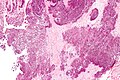
Pseudogout - intermed. mag. (WC)
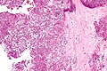
Pseudogout - high mag. (WC)


www:
Sign-out
KNEE - BONE AND SOFT TISSUE, RIGHT, KNEE ARTHROPLASTY:
- DEGENERATIVE JOINT DISEASE WITH SYNOVIAL HYPERPLASIA AND
  NO SIGNIFICANT INFLAMMATION.
- CRYSTALLINE DEPOSITS CONSISTENT WITH PSEUDOGOUT.
- BONE WITH THIN TRABECULAE.
Micro
The soft tissue section shows readily apparent rhomboid-shaped crystalline deposits (compatible with pseudogout). The crystals polarize and have a light blue hue under polarized light. Synovial hyperplasia is present. No lymphoid aggregates are apparent.
The bony section show thin bony trabeculae and cartilage with degenerative changes (surface fibrillation, thinning).
See also
References
- ↑ Yeung, J.C.; Leonard, Blair J. N. (2005). The Toronto Notes 2005 - Review for the MCCQE and Comprehensive Medical Reference (2005 ed.). The Toronto Notes Inc. for Medical Students Inc.. pp. RH6. ISBN 978-0968592854.
- ↑ WG. 8 January 2010.
- ↑ Saukko, Pekka; Knight, Bernard (2004). Knight's Forensic Pathology (3rd ed.). A Hodder Arnold Publication. pp. 589. ISBN 978-0340760444.
- ↑ URL: http://www.ncbi.nlm.nih.gov/pubmedhealth/PMH0001458/. Accessed on: 28 October 2011.
- ↑ Abhishek, A.; Doherty, S.; Maciewicz, R.; Muir, K.; Zhang, W.; Doherty, M. (Aug 2013). "Association between low cortical bone mineral density, soft-tissue calcification, vascular calcification and chondrocalcinosis: a case-control study.". Ann Rheum Dis. doi:10.1136/annrheumdis-2013-203400. PMID 23912799.
- ↑ 6.0 6.1 URL: http://www.learningradiology.com/archives04/COW%20102-CPPD/cppdcorrectpage.htm. Accessed on: 9 October 2013.
- ↑ Dieppe, P.; Swan, A. (May 1999). "Identification of crystals in synovial fluid.". Ann Rheum Dis 58 (5): 261-3. PMC 1752883. PMID 10225806. https://www.ncbi.nlm.nih.gov/pmc/articles/PMC1752883/.