Difference between revisions of "Cortical tuber"
Jump to navigation
Jump to search
Jensflorian (talk | contribs) (GFAP image added) |
Jensflorian (talk | contribs) (→General: link) |
||
| Line 14: | Line 14: | ||
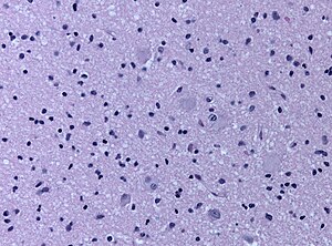
File:Tuber_GFAP_20191014_012.jpg|GFAP sparing dysmorphic ballon cells. | File:Tuber_GFAP_20191014_012.jpg|GFAP sparing dysmorphic ballon cells. | ||
</gallery> | </gallery> | ||
==Imaging== | |||
Examples on Radiopedia [[https://radiopaedia.org/articles/cortical-tubers]] | |||
==DDx== | ==DDx== | ||
Revision as of 13:07, 14 October 2019

Cortical tubers are malformative lesions in the CNS observed in tuberous sclerosis complex (abbreviated TSC), an autosomal dominant syndrome.
General
- Cortical tubers are malformative, epilepsy-associated.[1]
- Seen in 80-90% of the TSC cases.
- Gyrus is usu. thickened, raised, and occasionally dimpled.
- Giant cells, dysmorphic neurons, disrupted cortical lamination, gliosis, calcifications.
- Ballon cells are Vim+ve, MAP2+ve, Nestin+ve, GFAP+/-ve, NeuN+/-ve.
- TSC2 has larger and more numerous tubers.[2]
GFAP sparing dysmorphic ballon cells.

Imaging
Examples on Radiopedia [[1]]
DDx
- Focal cortical dysplasia ILAE type IIB (Tubers are usu. multifocal).
See also
References
- ↑ Cotter, JA. (Apr 2019). "An update on the central nervous system manifestations of tuberous sclerosis complex.". Acta Neuropathol. doi:10.1007/s00401-019-02003-1. PMID 30976976.
- ↑ Overwater, IE.; Swenker, R.; van der Ende, EL.; Hanemaayer, KB.; Hoogeveen-Westerveld, M.; van Eeghen, AM.; Lequin, MH.; van den Ouweland, AM. et al. (12 2016). "Genotype and brain pathology phenotype in children with tuberous sclerosis complex.". Eur J Hum Genet 24 (12): 1688-1695. doi:10.1038/ejhg.2016.85. PMID 27406250.