Difference between revisions of "Compound nevus"
| Line 5: | Line 5: | ||
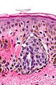
| Caption = Compound nevus. [[H&E stain]]. | | Caption = Compound nevus. [[H&E stain]]. | ||
| Synonyms = compound melanocytic nevus | | Synonyms = compound melanocytic nevus | ||
| Micro = nests of melanocytes, melanocytes "mature" with depth, usu. no mitoses (occ. superficial), no destruction of surrounding structures, no conspicuous nucleoli, | | Micro = nests of melanocytes in dermis ''and'' epidermis, melanocytes "mature" with depth, usu. no mitoses (occ. superficial), no destruction of surrounding structures, no conspicuous nucleoli, no significant melanocyte enlargement | ||
| Subtypes = | | Subtypes = | ||
| LMDDx = [[malignant melanoma]] (nevoid), [[junctional nevus]], [[intradermal nevus]], [[dysplastic nevus]] | | LMDDx = [[malignant melanoma]] (nevoid), [[junctional nevus]], [[intradermal nevus]], [[dysplastic nevus]] | ||
Revision as of 04:47, 25 January 2014
| Compound nevus | |
|---|---|
| Diagnosis in short | |
 Compound nevus. H&E stain. | |
|
| |
| Synonyms | compound melanocytic nevus |
|
| |
| LM | nests of melanocytes in dermis and epidermis, melanocytes "mature" with depth, usu. no mitoses (occ. superficial), no destruction of surrounding structures, no conspicuous nucleoli, no significant melanocyte enlargement |
| LM DDx | malignant melanoma (nevoid), junctional nevus, intradermal nevus, dysplastic nevus |
| Gross | pigment skin lesion, usu. small, regular border, no irregularity in pigmentation |
| Site | skin - see melanocytic lesions and common nevus |
|
| |
| Prevalence | very common |
| Prognosis | benign |
| Clin. DDx | pigmented skin lesions |
Compound nevus (abbreviated CN), also compound melanocytic nevus (abbreviated CMN), is a common benign melanocytic lesion.
The compound nevus is in the large group common nevus. In common language, nevus is known as a mole.
General
- Benign.
- Common.
- Think melanoma.
Clinical:
- ABCD = asymmetric, borders (irregular), colour (black), diameter (large).
Microscopic
Features:
- Symmetrical lesion.
- "Matures" with depth.
- Less cellular with depth.
- Less nuclear atypia with depth.
- Smaller cells with depth.
- Smaller nests with depth.
- Rare mitoses (superficial).
- No deep mitoses.
- No destruction of surrounding structures.
- No nucleoli.
- In the dermis and epidermis - key feature.
- +/-Adipocytes - uncommon.[1]
Images
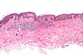
CN - low mag.
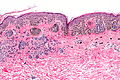
CN - intermed. mag.
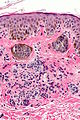
CN - high mag.
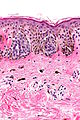
CN - high mag.
CN - very high mag.


Sign out
SKIN LESION, RIGHT NECK, SHAVE BIOPSY:
- BENIGN COMPOUND NEVUS.
Micro
The sections show melanocytes in the dermis and epidermis. The lesion is symmetrical in its architecture and pigment distribution. There is no pagetoid spread of melanocytes in the epidermis. Superficially, melanocytes are in nests. Melanocytes show no apparent cytologic atypia and mature with depth. No mitotic activity is appreciated. The lesion is completely excised in the plane of section.
Barely compound
The sections show melanocytes predominantly in the dermis. Rare melanocytic nests are seen at the dermal-epidermal junction. There is no pagetoid spread of melanocytes in the epidermis. The lesion is symmetrical in its architecture. Superficially, melanocytes are in nests and pigment is present. The melanocytes mature with depth. No mitotic activity is appreciated. The lesion is present at the margin.
Congenital features present
The sections show melanocytes in the dermis and epidermis. There is no pagetoid spread of melanocytes in the epidermis. Superficially, melanocytes are in nests. Melanocytes show mild nuclear enlargement but mature with depth. Nucleoli are not prominent. Melanocytes are found adjacent to adnexal structures and blood vessels. No mitotic activity is appreciated. The lesion is incompletely excised.