Complex endometrial hyperplasia
| Complex endometrial hyperplasia | |
|---|---|
| Diagnosis in short | |
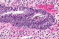
 Complex endometrial hyperplasia. H&E stain. | |
|
| |
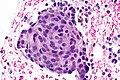
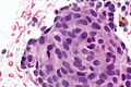
| LM | increase in size & number of glands + irregular shape, nuclear pseudostratification, nuclear enlargement, mitoses common, +/-nuclear atypia (round nuclei ~ 2-3x the size of a lymphocyte, grey/translucent chromatin, nucleoli) |
| Subtypes | CEH NOS (CEH without atypia), CEH with atypia |
| LM DDx | endometrioid endometrial carcinoma, artifactual gland crowding |
| Site | endometrium - see endometrial hyperplasia |
|
| |
| Associated Dx | obesity |
| Clinical history | usu. premenopausal or perimenopausal |
| Symptoms | abnormal uterine bleeding (AUB) |
| Prevalence | common |
| Clin. DDx | dysfunctional uterine bleeding (diagnosis of exclusion), endometrial carcinoma, atrophy, endometrial polyp, others |
| Complex endometrial hyperplasia | |
|---|---|
| External resources | |
| EHVSC | 10169 10181 (CEH with atypia) |
Complex endometrial hyperplasia, abbreviated CEH, is a relatively common pre-malignant pathology of the endometrium.
It is generally subdivided into complex endometrial hyperplasia without atypia and complex endometrial hyperplasia with atypia.
CEH with atypia is also known as complex atypical hyperplasia.
General
- Usually premenopausal or perimenopausal women.
- Associated with obesity.
- High risk of transformation to endometrial carcinoma especially when with atypia.
Microscopic
Features:
- Increase in size & number of glands + irregular shape - key feature.
- Cell stratification.
- Nuclear enlargement.
- Mitoses common.
- +/-Nuclear atypia:
- Round nuclei ~ 2-3x the size of a lymphocyte.
- Grey/translucent chromatin.
- Nucleoli.
Notes:
- Normal "gland-to-stroma ratio" is 1:3.
- Two "touching" glands may be one gland in section.
- Atypical nuclei often hide between non-typical nuclei, like peg cells in the fallopian tube.
DDx:
- Artifactual gland crowding.
- Endometrioid endometrial carcinoma - see endometrial carcinoma versus complex endometrial hyperplasia.
Endometrial carcinoma versus complex endometrial hyperplasia
Complex endometrial hyperplasia:
- Non-confluent - glands distinct from one another.
Classic criteria for endometrial carcinoma
This is pimping material that shows up on exams.
Endometrial carcinoma has one of the following:[1][2][3]
- Desmoplastic stromal response.
- Confluent cribriform growth. †
- Extensive papillary growth. †
- Severe cytologic atypia. †
Note:
- † There is a size cut-off for criteria 2, 3 and 4: > 2.1 mm.[2]
How to remember ABCDE:
- Atypia Bad.
- Confluent cribriform growth.
- Desmoplasia.
- Extensive papillary growth.
Images
Complex endometrial hyperplasia:
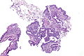
CEH - low mag.
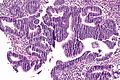
CEH - intermed. mag.
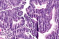
CEH - high mag.


Squamous morules - commonly associated with hyperplasia and malignancy:
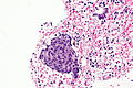
SM - high mag.
SM - very high mag.
SM - extremely high mag.


Fused glands suggestive of CEH in a polyp:
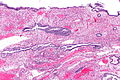
Endometrial polyp with fused glands - low mag. (WC)
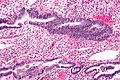
Endometrial polyp with fused glands - intermed. mag. (WC)
Endometrial polyp with fused glands - high mag. (WC)


www:
- Endometrial hyperplasia (webpathology.com).
- Complex endometrial hyperplasia with atypia (webpathology.com).
Sign out
CEH without atypia
ENDOMETRIUM, BIOPSY:
- COMPLEX ENDOMETRIAL HYPERPLASIA.
-- NEGATIVE FOR CYTOLOGIC ATYPIA.
ENDOMETRIUM, BIOPSY:
- SMALL FOCUS OF COMPLEX ENDOMETRIAL HYPERPLASIA WITHOUT ATYPIA, WITH
  SQUAMOUS MORULES.
- ENDOMETRIAL POLYP WITH ONE ATYPICAL GLAND AND A SQUAMOUS MORULE.
- SCANT ENDOCERVICAL EPITHELIUM WITHOUT APPARENT PATHOLOGY.
CEH with atypia
Insufficient confluence for carcinoma
ENDOMETRIUM, BIOPSY:
- COMPLEX ENDOMETRIAL HYPERPLASIA WITH ATYPIA, SEE COMMENT.

COMMENT:
The sections show architecturally complex crowded glands with focal
morular squamous metaplasia and focal cribriforming.  Desmoplasia
is not identified. The degree of gland confluence is not considered
sufficient for the diagnosis of endometrial carcinoma. Nuclear atypia
is present focally.
Insufficient extent for carcinoma
ENDOMETRIUM, BIOPSY:
- COMPLEX ENDOMETRIAL HYPERPLASIA WITH ATYPIA, SEE COMMENT.

COMMENT:
The sections show architecturally complex back-to-back glands with focal
morular squamous metaplasia and cribriforming. Desmoplasia is not present.
The extent, i.e. the size of the abnormality, is not considered sufficient
for the diagnosis of endometrial carcinoma.
See also
References
- ↑ Nucci, Marisa R.; Oliva, Esther (2009). Gynecologic Pathology: A Volume in Foundations in Diagnostic Pathology Series (1st ed.). Churchill Livingstone. pp. 239. ISBN 978-0443069208.
- ↑ 2.0 2.1 Kurman, RJ.; Norris, HJ. (Jun 1982). "Evaluation of criteria for distinguishing atypical endometrial hyperplasia from well-differentiated carcinoma.". Cancer 49 (12): 2547-59. PMID 7074572.
- ↑ URL: http://www.cap.org/apps/docs/committees/cancer/cancer_protocols/2011/Endometrium_11protocol.pdf. Accessed on: 12 January 2012.