Coffee bean nucleus
Jump to navigation
Jump to search
The printable version is no longer supported and may have rendering errors. Please update your browser bookmarks and please use the default browser print function instead.


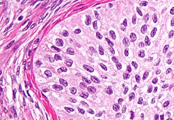
Coffee bean nucleus is a cell nucleus that looks like a coffee bean. Coffee bean nuclei have a limited differential diagnosis.
Microscopic
Features - coffee bean nuclei have:
- An ellipsoid-shape.
- A central groove (along the long axis of the nucleus).
Images
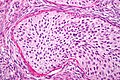
Coffee bean nuclei in a Brenner tumour (WC)

Classic differential diagnosis
Others considerations
- Invasive ductal carcinoma of the pancreas.
- Langerhans cell histiocytosis.[3]
- Papillary thyroid carcinoma. (???)
- Walthard cell rest.
See also
References
- ↑ Vodovnik, A. (Jun 2002). "Bladder-washing cytology of metastatic ovarian granulosa cell tumor.". Diagn Cytopathol 26 (6): 387-8. doi:10.1002/dc.10095. PMID 12112830.
- ↑ Ahr, A.; Arnold, G.; Göhring, UJ.; Costa, S.; Scharl, A.; Gauwerky, JF.. "Cytology of ascitic fluid in a patient with metastasizing malignant Brenner tumor of the ovary. A case report.". Acta Cytol 41 (4 Suppl): 1299-304. PMID 9990262.
- ↑ Yap, WM.; Chuah, KL.; Tan, PH. (Feb 2001). "Langerhans cell histiocytosis involving the thyroid and parathyroid glands.". Mod Pathol 14 (2): 111-5. doi:10.1038/modpathol.3880266. PMID 11235902.