Difference between revisions of "Coffee bean nucleus"
Jump to navigation
Jump to search
| Line 13: | Line 13: | ||
==Classic differential diagnosis== | ==Classic differential diagnosis== | ||
*[[Granulosa cell tumour]]. | *[[Granulosa cell tumour]]. | ||
*[[Brenner tumour]]. | *[[Brenner tumour]].<ref>{{Cite journal | last1 = Ahr | first1 = A. | last2 = Arnold | first2 = G. | last3 = Göhring | first3 = UJ. | last4 = Costa | first4 = S. | last5 = Scharl | first5 = A. | last6 = Gauwerky | first6 = JF. | title = Cytology of ascitic fluid in a patient with metastasizing malignant Brenner tumor of the ovary. A case report. | journal = Acta Cytol | volume = 41 | issue = 4 Suppl | pages = 1299-304 | month = | year = | doi = | PMID = 9990262 }}</ref> | ||
===Others considerations=== | ===Others considerations=== | ||
Revision as of 19:04, 30 September 2014


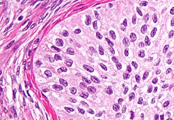
Coffee bean nucleus is a cell nucleus that looks like a coffee bean. Coffee bean nuclei have a limited differential diagnosis.
Microscopic
- Coffee bean nuclei are ellipsoid and have a central groove.
Images
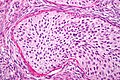
Coffee bean nuclei in a Brenner tumour (WC)

Classic differential diagnosis
Others considerations
- Invasive ductal carcinoma of the pancreas.
- Langerhans cell histiocytosis.
- Papillary thyroid carcinoma. (???)