Difference between revisions of "Central neurocytoma"
Jump to navigation
Jump to search
(redirect, +cat) |
(split-out) |
||
| Line 1: | Line 1: | ||
'''Central neurocytoma''', abbreviated '''CNC''', is a rare [[neuropathology tumour]]. | |||
==General== | |||
*Rare - less than 1% of brain tumours.<ref name=pmid16163043>{{Cite journal | last1 = Chuang | first1 = MT. | last2 = Lin | first2 = WC. | last3 = Tsai | first3 = HY. | last4 = Liu | first4 = GC. | last5 = Hu | first5 = SW. | last6 = Chiang | first6 = IC. | title = 3-T proton magnetic resonance spectroscopy of central neurocytoma: 3 case reports and review of the literature. | journal = J Comput Assist Tomogr | volume = 29 | issue = 5 | pages = 683-8 | month = | year = | doi = | PMID = 16163043 }}</ref> | |||
*Benign. | |||
*First described in 1982.<ref name=pmid16163043/> | |||
==Gross/radiology== | |||
*Intraventricular.<ref>URL: [http://moon.ouhsc.edu/kfung/jty1/Com/Com307-1-Diss.htm http://moon.ouhsc.edu/kfung/jty1/Com/Com307-1-Diss.htm]. Accessed on: 12 January 2012.</ref> | |||
**Characteristically attached to the ''septum pellucidum''.<ref name=pmid20692674>{{Cite journal | last1 = Kerkeni | first1 = A. | last2 = Ben Lakhdher | first2 = Z. | last3 = Rkhami | first3 = M. | last4 = Sebai | first4 = R. | last5 = Belguith | first5 = L. | last6 = Khaldi | first6 = M. | last7 = Ben Hamouda | first7 = M. | title = [Central neurocytoma: Study of 32 cases and review of the literature]. | journal = Neurochirurgie | volume = 56 | issue = 5 | pages = 408-14 | month = Oct | year = 2010 | doi = 10.1016/j.neuchi.2010.07.001 | PMID = 20692674 }}</ref> | |||
==Microscopic== | |||
Features:<ref>URL: [http://moon.ouhsc.edu/kfung/jty1/Com/Com307-1-Diss.htm http://moon.ouhsc.edu/kfung/jty1/Com/Com307-1-Diss.htm]. Accessed on: 27 May 2011.</ref> | |||
*Perivascular pseudorosette = circular/flower-like arrangement of cells with blood vessel at the centre.<ref name=pmid16551982>{{cite journal |author=Wippold FJ, Perry A |title=Neuropathology for the neuroradiologist: rosettes and pseudorosettes |journal=AJNR Am J Neuroradiol |volume=27 |issue=3 |pages=488–92 |year=2006 |month=March |pmid=16551982 |doi= |url=}}</ref> | |||
*Islands of neuropil. | |||
*Polygonal cells with a perinuclear halo. | |||
DDx: | |||
*[[Oligodendroglioma]]. | |||
DDx of perivascular pseudorosette: | |||
*Ependymoma. | |||
*[[Medulloblastoma]], PNET. | |||
*[[Glioblastoma]]s. | |||
===Images=== | |||
<gallery> | |||
Image:Central_neurocytoma_-_intermed_mag.jpg | Central neurocytoma - intermed. mag. (WC) | |||
Image:Central_neurocytoma_-_high_mag.jpg | Central neurocytoma - oligodendrogllioma-like area - high mag. (WC) | |||
Image:Central_neurocytoma_-_2_-_high_mag.jpg | Central neurocytoma - pseudorosettes - high mag. (WC) | |||
Image:Central_neurocytoma_-_2_-_very_high_mag.jpg | Central neurocytoma - pseudorossettes - very high mag. (WC) | |||
</gallery> | |||
[[www]]: | |||
*[http://moon.ouhsc.edu/kfung/jty1/Com/Com307-1-Diss.htm Central neurocytoma - several images (ouhsc.edu)]. | |||
*[http://frontalcortex.com/?page=image&topic=1&qid=1203 Central neurocytoma (frontalcortex.com)]. | |||
*[http://path.upmc.edu/cases/case74/micro.html Central neurocytoma - crappy images (upmc.edu)]. | |||
==IHC== | |||
*MIB1 - high may predict re-occurance.<ref name=pmid15015671>{{Cite journal | last1 = Schmidt | first1 = MH. | last2 = Gottfried | first2 = ON. | last3 = von Koch | first3 = CS. | last4 = Chang | first4 = SM. | last5 = McDermott | first5 = MW. | title = Central neurocytoma: a review. | journal = J Neurooncol | volume = 66 | issue = 3 | pages = 377-84 | month = Feb | year = 2004 | doi = | PMID = 15015671 }}</ref> | |||
==See also== | |||
*[[Neuropathology tumours]]. | |||
==References== | |||
{{Reflist|2}} | |||
[[Category:Diagnosis]] | [[Category:Diagnosis]] | ||
[[Category:Neuropathology tumours]] | |||
Latest revision as of 06:24, 10 December 2014
Central neurocytoma, abbreviated CNC, is a rare neuropathology tumour.
General
Gross/radiology
Microscopic
Features:[4]
- Perivascular pseudorosette = circular/flower-like arrangement of cells with blood vessel at the centre.[5]
- Islands of neuropil.
- Polygonal cells with a perinuclear halo.
DDx:
DDx of perivascular pseudorosette:
- Ependymoma.
- Medulloblastoma, PNET.
- Glioblastomas.
Images
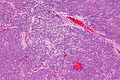
Central neurocytoma - intermed. mag. (WC)
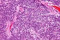
Central neurocytoma - oligodendrogllioma-like area - high mag. (WC)
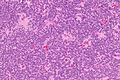
Central neurocytoma - pseudorosettes - high mag. (WC)
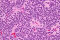
Central neurocytoma - pseudorossettes - very high mag. (WC)


www:
- Central neurocytoma - several images (ouhsc.edu).
- Central neurocytoma (frontalcortex.com).
- Central neurocytoma - crappy images (upmc.edu).
IHC
- MIB1 - high may predict re-occurance.[6]
See also
References
- ↑ 1.0 1.1 Chuang, MT.; Lin, WC.; Tsai, HY.; Liu, GC.; Hu, SW.; Chiang, IC.. "3-T proton magnetic resonance spectroscopy of central neurocytoma: 3 case reports and review of the literature.". J Comput Assist Tomogr 29 (5): 683-8. PMID 16163043.
- ↑ URL: http://moon.ouhsc.edu/kfung/jty1/Com/Com307-1-Diss.htm. Accessed on: 12 January 2012.
- ↑ Kerkeni, A.; Ben Lakhdher, Z.; Rkhami, M.; Sebai, R.; Belguith, L.; Khaldi, M.; Ben Hamouda, M. (Oct 2010). "[Central neurocytoma: Study of 32 cases and review of the literature].". Neurochirurgie 56 (5): 408-14. doi:10.1016/j.neuchi.2010.07.001. PMID 20692674.
- ↑ URL: http://moon.ouhsc.edu/kfung/jty1/Com/Com307-1-Diss.htm. Accessed on: 27 May 2011.
- ↑ Wippold FJ, Perry A (March 2006). "Neuropathology for the neuroradiologist: rosettes and pseudorosettes". AJNR Am J Neuroradiol 27 (3): 488–92. PMID 16551982.
- ↑ Schmidt, MH.; Gottfried, ON.; von Koch, CS.; Chang, SM.; McDermott, MW. (Feb 2004). "Central neurocytoma: a review.". J Neurooncol 66 (3): 377-84. PMID 15015671.