Difference between revisions of "Castleman disease"
Jump to navigation
Jump to search
(split out) |
|||
| Line 1: | Line 1: | ||
{{ Infobox diagnosis | |||
| Name = {{PAGENAME}} | |||
| Image = Castleman_disease_-_high_mag.jpg | |||
| Width = | |||
| Caption = Castleman disease (hyaline-vascular variant). [[H&E stain]]. | |||
| Synonyms = | |||
| Micro = | |||
| Subtypes = hyaline-vascular variant (HVV), plasma cell variant (PCV) | |||
| LMDDx = HVV: [[mantle cell lymphoma]] | |||
| Stains = | |||
| IHC = HVV: cyclin D1 -ve, other stains to exclude lymphoma; PCV: HHV-8 +ve | |||
| EM = | |||
| Molecular = | |||
| IF = | |||
| Gross = | |||
| Grossing = | |||
| Site = [[lymph node]] - see ''[[lymph node pathology]]'' | |||
| Assdx = | |||
| Syndromes = | |||
| Clinicalhx = | |||
| Signs = | |||
| Symptoms = | |||
| Prevalence = rare | |||
| Bloodwork = | |||
| Rads = | |||
| Endoscopy = | |||
| Prognosis = | |||
| Other = | |||
| ClinDDx = | |||
| Tx = | |||
}} | |||
'''Castleman disease''', abbreviated '''CD''', is a rare [[Lymph node pathology|pathology of the lymph node]]. | '''Castleman disease''', abbreviated '''CD''', is a rare [[Lymph node pathology|pathology of the lymph node]]. | ||
| Line 5: | Line 36: | ||
==General== | ==General== | ||
*Benign. | *Benign. | ||
*Hyaline vascular variant - a pathology of the follicular dendritic cells.<ref>{{Cite journal | last1 = Cokelaere | first1 = K. | last2 = Debiec-Rychter | first2 = M. | last3 = De Wolf-Peeters | first3 = C. | last4 = Hagemeijer | first4 = A. | last5 = Sciot | first5 = R. | title = Hyaline vascular Castleman's disease with HMGIC rearrangement in follicular dendritic cells: molecular evidence of mesenchymal tumorigenesis. | journal = Am J Surg Pathol | volume = 26 | issue = 5 | pages = 662-9 | month = May | year = 2002 | doi = | PMID = 11979097 }}</ref> | *Hyaline vascular variant (classic Castleman disease) - a pathology of the follicular dendritic cells.<ref>{{Cite journal | last1 = Cokelaere | first1 = K. | last2 = Debiec-Rychter | first2 = M. | last3 = De Wolf-Peeters | first3 = C. | last4 = Hagemeijer | first4 = A. | last5 = Sciot | first5 = R. | title = Hyaline vascular Castleman's disease with HMGIC rearrangement in follicular dendritic cells: molecular evidence of mesenchymal tumorigenesis. | journal = Am J Surg Pathol | volume = 26 | issue = 5 | pages = 662-9 | month = May | year = 2002 | doi = | PMID = 11979097 }}</ref> | ||
===Classification=== | ===Classification=== | ||
Revision as of 01:43, 26 December 2013
| Castleman disease | |
|---|---|
| Diagnosis in short | |
 Castleman disease (hyaline-vascular variant). H&E stain. | |
| Subtypes | hyaline-vascular variant (HVV), plasma cell variant (PCV) |
| LM DDx | HVV: mantle cell lymphoma |
| IHC | HVV: cyclin D1 -ve, other stains to exclude lymphoma; PCV: HHV-8 +ve |
| Site | lymph node - see lymph node pathology |
|
| |
| Prevalence | rare |
Castleman disease, abbreviated CD, is a rare pathology of the lymph node.
It is also known as angiofollicular lymph node hyperplasia and giant lymph node hyperplasia.[1]
General
- Benign.
- Hyaline vascular variant (classic Castleman disease) - a pathology of the follicular dendritic cells.[2]
Classification
CD is grouped by histologic appearance:[3]
- Hyaline vascular (HV) variant (described by Castleman).
- Usually unicentric.
- Typically mediastinal or axial.
- More common than plasma cell variant; represents 80-90% of CD cases.
- May be associated with follicular dendritic cell neoplasia.[4]
- Plasma cell (PC) variant.
- Usually multicentric, may be unicentric.
- Abundant plasma cells.
- Associated with HHV-8 infection (the same virus implicated in Kaposi's sarcoma).
Notes:
- The subclassification of CD is in some flux. Some authors advocate splitting-out HHV-8 and multicentric as separate subtypes.[5]
Microscopic
Hyaline-vascular variant
- Pale concentric (expanded) mantle zone lymphocytes - key feature.
- "Regressed follicles" - germinal center (pale area) is small.
- "Lollipops":
- Germinal centers fed by prominent (radially penetrating sclerotic) vessels; lollipop-like appearance.
- Two germinal centers in one follicle.
- Hyaline material (pink acellular stuff on H&E) in germinal center.
- Sinuses effaced (lost).
- Mitoses absent.
Images
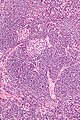
CD HVV - "lollipop" sign - high mag. (WC)
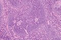
CD HVV - showing expanded mantle zone - intermed. mag. (WC)

www:
Plasma cell variant
Features:[7]
- Interfollicular sheets of plasma cells - key feature.
- Active germinal centers - mitoses present.
- Sinus perserved.
IHC
Hyaline-vascular variant:
- Stains to exclude mantle cell lymphoma:
- Cyclin D1.
Plasma cell variant:
- HHV-8 +ve.
See also
References
- ↑ URL: http://www.mayoclinic.com/health/castleman-disease/DS01000. Accessed on: 17 June 2010.
- ↑ Cokelaere, K.; Debiec-Rychter, M.; De Wolf-Peeters, C.; Hagemeijer, A.; Sciot, R. (May 2002). "Hyaline vascular Castleman's disease with HMGIC rearrangement in follicular dendritic cells: molecular evidence of mesenchymal tumorigenesis.". Am J Surg Pathol 26 (5): 662-9. PMID 11979097.
- ↑ Ioachim, Harry L; Medeiros, L. Jeffrey (2008). Ioachim's Lymph Node Pathology (4th ed.). Lippincott Williams & Wilkins. pp. 228. ISBN 978-0781775960.
- ↑ Humphrey, Peter A; Dehner, Louis P; Pfeifer, John D (2008). The Washington Manual of Surgical Pathology (1st ed.). Lippincott Williams & Wilkins. pp. 596. ISBN 978-0781765275.
- ↑ Cronin, DM.; Warnke, RA. (Jul 2009). "Castleman disease: an update on classification and the spectrum of associated lesions.". Adv Anat Pathol 16 (4): 236-46. doi:10.1097/PAP.0b013e3181a9d4d3. PMID 19546611.
- ↑ URL: http://www.ispub.com/journal/the_internet_journal_of_otorhinolaryngology/volume_9_number_2_11/article/a_rare_case_of_castleman_s_disease_presenting_as_cervical_neck_mass.html. Accessed on: 15 June 2010.
- ↑ 7.0 7.1 Ioachim, Harry L; Medeiros, L. Jeffrey (2008). Ioachim's Lymph Node Pathology (4th ed.). Lippincott Williams & Wilkins. pp. 236. ISBN 978-0781775960.